

Case Report: Unusual Heterotopic Ossification of the Hindfoot
- PMID: 35747430
- PMCID: PMC9209653
- DOI: 10.3389/fsurg.2022.917560
Case Report: Unusual Heterotopic Ossification of the Hindfoot
Abstract
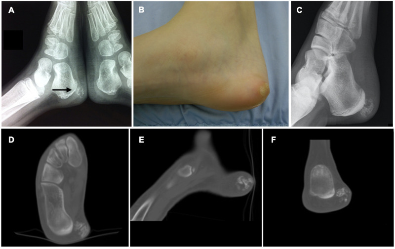
Heterotopic ossification (HO) is a pathologic condition in which aberrant lamellar bone deposits in soft tissues, outside of the normal skeleton. Pathogenesis is still unclear, but different risk factors are known. Here we report a case of a 14 year-old girl presenting with pain in the medial calcaneal region and evidence of a rapidly growing, firm and solid neoformation. The lesion was diagnosed 6 years earlier, but it was consistently smaller and asymptomatic so that the patient did not undergo any follow up. The patient had no previous trauma or surgery, no other risk factors for HO and did not show any clinically evident HO in other districts. Xray and CT showed a heterogeneous bony lesion in the context of soft tissues, isolated from the calcaneus. After complete excision, histological analysis confirmed the diagnosis of HO. In conclusion, lone non congenital HO can occur regardless of known risk factors. Small HO lesion may also enter a proliferative phase without evidence of triggering events. More studies are required to better understand etiopathogenesis of HO in these clinical settings.
Keywords: bone; case report; children; foot; heterotopic ossification.
Copyright © 2022 Danya, Marco, Valentino, Mario and Antonio Pompilio.
Conflict of interest statement
The authors declare that the research was conducted in the absence of any commercial or financial relationships that could be construed as a potential conflict of interest..
Figures


References
-
- Riegler HF, Harris CM. Heterotopic bone formation after total hip arthroplasty. - Cerca con Google. https://www.google.com/search?q=Riegler+HF%2C+Harris+CM.+Heterotopic+bon... (Accessed December 17, 2020). - PubMed
Publication types
LinkOut - more resources
Full Text Sources