Case Reports
. 2022 Jul;12(3):520-523.
doi: 10.1177/19418744221079145. Epub 2022 Mar 4.

COVID-19-associated Isolated Cortical Vein Thrombosis: Detection of SARS-CoV-2 in CSF

Affiliations
Case Reports

COVID-19-associated Isolated Cortical Vein Thrombosis: Detection of SARS-CoV-2 in CSF

Ayla Çulha Oktar et al. Neurohospitalist. 2022 Jul.

Abstract

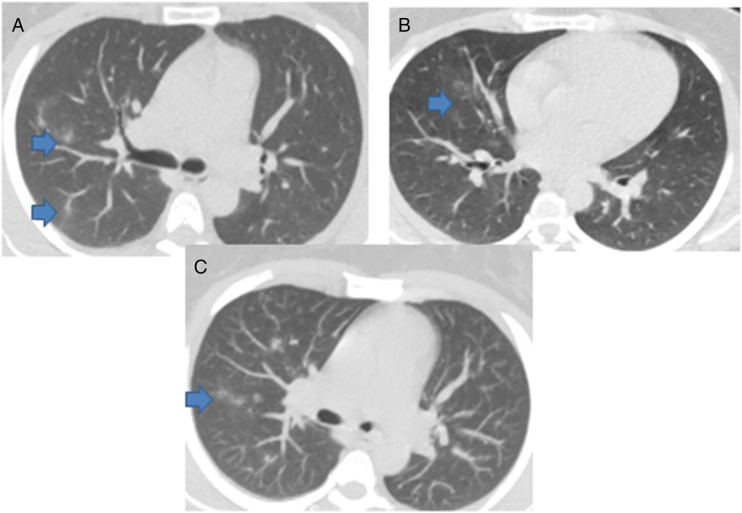
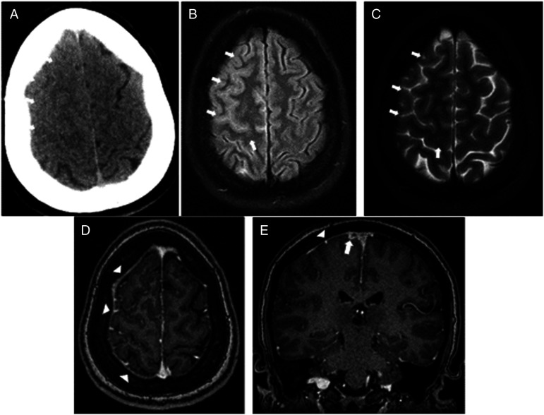
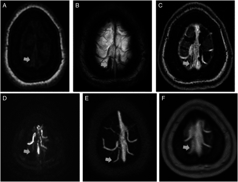
COVID-19 has been associated with central nervous system manifestations; however, cerebral venous thrombosis is rarely reported. A 34-year-old woman was admitted to the hospital with headache and recurrent seizures; she was recently discharged after COVID-19 pneumonia. Cranial magnetic resonance imaging and magnetic resonance venography showed cortical vein thrombosis in the right frontal lobe. SARS-CoV-2 RNA was detected in cerebrospinal fluid analysis. The patient was anticoagulated and put on antiepileptics. The most probable mechanism underlying the venous thrombosis is COVID-19-associated hypercoagulability. However, the relation between the viral RNA in cerebrospinal fluid analysis and the thrombosis is controversial.

Keywords: COVID-19; intracranial sinus thrombosis; seizures.


Conflict of interest statement

Declaration of Conflicting Interests: The author(s) declared no potential conflicts of interest with respect to the research, authorship, and/or publication of this article.

Figures

Figure 1.
Figure 1.
(A-B) Non-enhanced axial chest CT and (C) axial MIP images show unilateral peripheral and central, solid ground glass mix type nodular opacities of right upper, lower, middle lobes (arrows).
Figure 2.
Figure 2.
Demonstration of sulcal effacement of right frontal cerebral sulci at the level of vertex and unsuppressed CSF signal representing the meningeal irritation (short arrows). (A) Non-enhanced axial head CT, (B) axial FLAIR, (C) axial T2-WI (D) contrasted volumetric axial T1-WI shows mild dural enhancement (arrowheads), and (E) contrasted MPR coronal T1-WI shows dural enhancement (arrowhead) and filling defect of cortical vein (short arrow).
Figure 3.
Figure 3.
(A) Non-enhanced axial T1-WI, (B) axial SWI MIP, (C) contrasted T1-WI, (D) phase contrast MRV axial MIP reconstruction show thrombosed right cortical vein, and (E and F) contrasted MRV and contrasted T1-WI (short arrows) demonstrate the partial filling defect of thrombosed cortical vein after medication

References

    1. Mao L, Jin H, Wang M, et al. Neurologic manifestations of hospitalized patients with Coronavirus disease 2019 in Wuhan, China. JAMA Neurol. 2020;77(6):683-690. doi:10.1001/jamaneurol.2020.1127. - DOI - PMC - PubMed
    1. Giacomelli A, Pezzati L, Conti F, et al. Self-reported olfactory and taste disorders in patients with severe acute respiratory Coronavirus 2 infection: A cross-sectional study. Clin Infect Dis. 2020;71(15):889-890. doi:10.1093/cid/ciaa330. - DOI - PMC - PubMed
    1. Klok FA, Kruip MJHA, van der Meer NJM, et al. Incidence of thrombotic complications in critically ill ICU patients with COVID-19. Thromb Res. 2020;191:145-147. doi:10.1016/j.thromres.2020.04.013. - DOI - PMC - PubMed
    1. Lewis A, Frontera J, Placantonakis DG, et al. Cerebrospinal fluid in COVID-19: A systematic review of the literature. J Neurol Sci. 2021;421:117316. doi:10.1016/j.jns.2021.117316. - DOI - PMC - PubMed
    1. Li Y, Li M, Wang M, et al. Acute cerebrovascular disease following COVID-19: A single center, retrospective, observational study. Stroke and Vascular Neurology. 2020;5:279-284. doi:10.1136/svn-2020-000431.Published online July 2, 2020:svn-2020-000431 - DOI - PMC - PubMed

Publication types