. 2022 Jun;5(3):274-280.
doi: 10.1002/ame2.12252. Epub 2022 Jun 23.

Distinct neuronal excitability alterations of medial prefrontal cortex in early-life neglect model of rats

Affiliations

Distinct neuronal excitability alterations of medial prefrontal cortex in early-life neglect model of rats

Yu Zhang et al. Animal Model Exp Med. 2022 Jun.

Abstract

Object: Early-life neglect has irreversible emotional effects on the central nervous system. In this work, we aimed to elucidate distinct functional neural changes in medial prefrontal cortex (mPFC) of model rats.

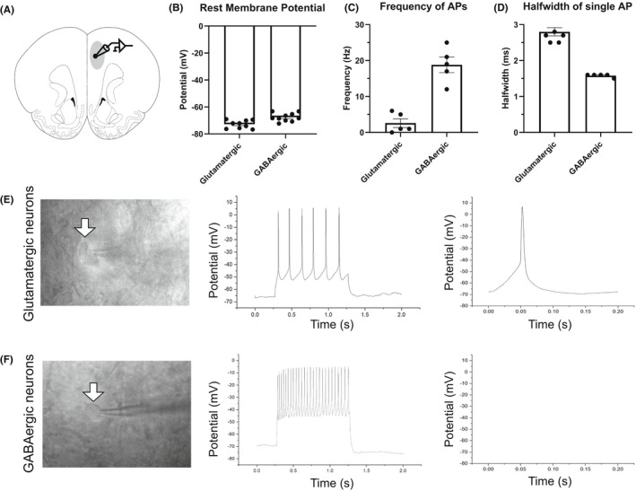
Methods: Maternal separation with early weaning was used as a rat model of early-life neglect. The excitation of glutamatergic and GABAergic neurons in rat mPFC was recorded and analyzed by whole-cell patch clamp.

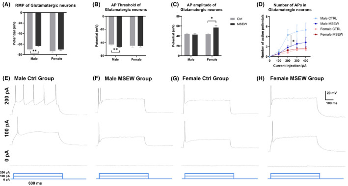
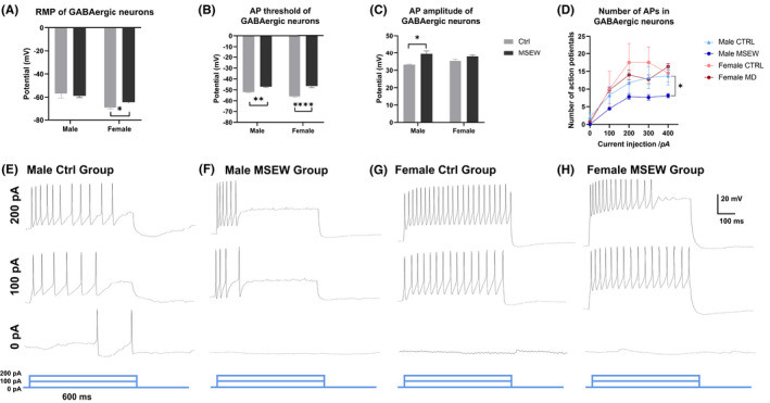
Results: Glutamatergic and GABAergic neurons of mPFC were distinguished by typical electrophysiological properties. The excitation of mPFC glutamatergic neurons was significantly increased in male groups, while the excitation of mPFC GABAergic neurons was significant in both female and male groups, but mainly in terms of rest membrane potential and amplitude, respectively.

Conclusions: Glutamatergic and GABAergic neurons in medial prefrontal cortex showed different excitability changes in a rat model of early-life neglect, which can contribute to distinct mechanisms for emotional and cognitive manifestations.

Keywords: GABAergic; early-life neglect model; glutamatergic; maternal separation with early weaning; medial prefrontal cortex; neuronal excitability.


Conflict of interest statement

Yu Zhang is an Editorial Board member of AMEM and a coauthor of this article. To minimize bias, they were excluded from all editorial decision‐making related to the acceptance of this article for publication. Chuan Qin is the Editor‐in‐Chief of AMEM and a co‐author of this article. They were excluded from editorial decision‐making related to the acceptance and publication of this article.

Figures

FIGURE 1
FIGURE 1
Electrophysiological properties of putative glutamatergic and GABAergic neurons in medial prefrontal cortex. (A) Schematic coronal section of a rat brain, showing the medial prefrontal cortex (mPFC). Recording electrodes were placed in layer II/III of mPFC. Excitation of neurons were recorded by whole‐cell patch clamp. (B–D) Summary histograms demonstrating different electrophysiological properties of mPFC glutamatergic and GABAergic neurons, regarding rest membrane potentials (B), action potential firing frequency (C), and halfwidth (D). (E) Typical example of a putative glutamatergic neuron from mPFC. Left, pyramidal shape of a mPFC glutamatergic neuron. Middle, multiple action potentials triggered by 1‐ms depolarizing current of 100 pA. Right, a typical single action potential from the neuron in left panel. (F) Typical example of a putative GABAergic neuron from mPFC. Left, oval shape of a mPFC GABAergic neuron. Middle, multiple action potentials triggered by 1 ms depolarizing current of 100 pA. Right, a single action potential from the neuron in left panel.
FIGURE 2
FIGURE 2
Distinct alterations in glutamatergic neuronal excitation between male and female rats. (A) Altered rest membrane potentials of mPFC glutamatergic neurons between male control and male MSEW group. (B) Altered threshold of a single action potential of mPFC glutamatergic neurons between male control and male MSEW group. (C) Increased amplitude of a single action potential of mPFC glutamatergic neurons in female MSEW group. (D) Multiple action potentials of 4 groups. Decreased number of APs between male groups. (E–H) Representative multiple action potentials of the 4 groups (*p < .05; **p < .01, MSEW group compared with control group. N = 10 cells for each group).
FIGURE 3
FIGURE 3
Distinct alterations in GABAergic neuronal excitation between male and female rats. (A) Altered rest membrane potentials of mPFC GABAergic neurons between female control and female MSEW group. (B) Altered threshold of a single action potential of mPFC GABAergic neurons between control and MSEW groups. (C) Increased amplitude of a single action potential of mPFC glutamatergic neurons in male MSEW group. (D) Multiple action potentials of 4 groups. Decreased number of APs in between male groups. (E–H) Representative multiple action potentials of the 4 groups (*p < .05; **p < .01; ****p < .0001, MSEW group compared with control group. N = 10 cells for each group).

References

    1. Matjasko JL, Herbst JH, Estefan LF. Preventing adverse childhood experiences: the role of etiological, evaluation, and implementation research. Am J Prev Med. 2022;62(6S1):S6‐S15. - PMC - PubMed
    1. Miguel PM, Pereira LO, Silveira PP, Meaney MJ. Early environmental influences on the development of children's brain structure and function. Dev Med Child Neurol. 2019;61(10):1127‐1133. - PubMed
    1. Sinani A, Vassi A, Tsotsokou G, Nikolakopoulou M, Kouvelas ED, Mitsacos A. Early life stress influences basal ganglia dopamine receptors and novel object recognition of adolescent and adult rats. IBRO Neurosci Rep. 2022;12:342‐354. - PMC - PubMed
    1. Baracz SJ, Robinson KJ, Wright AL, et al. Oxytocin as an adolescent treatment for methamphetamine addiction after early life stress in male and female rats. Neuropsychopharmacology. 2022. Epub ahead of print. 10.1038/s41386-022-01336-y - DOI - PMC - PubMed
    1. Sanchez EO, Bangasser DA. The effects of early life stress on impulsivity. Neurosci Biobehav Rev. 2022;137:104638. - PMC - PubMed

Publication types

LinkOut - more resources