. 2022 Jul;63(7):665-674.
doi: 10.3349/ymj.2022.63.7.665.

What Affects Segmental Lordosis of the Surgical Site after Minimally Invasive Transforaminal Lumbar Interbody Fusion?

Affiliations

What Affects Segmental Lordosis of the Surgical Site after Minimally Invasive Transforaminal Lumbar Interbody Fusion?

Soo-Heon Kim et al. Yonsei Med J. 2022 Jul.

Abstract

Purpose: This study was undertaken to identify factors that affect segmental lordosis (SL) after minimally invasive transforaminal lumbar interbody fusion (MIS-TLIF) by comparing patients whose postoperative SL increased with those whose decreased.

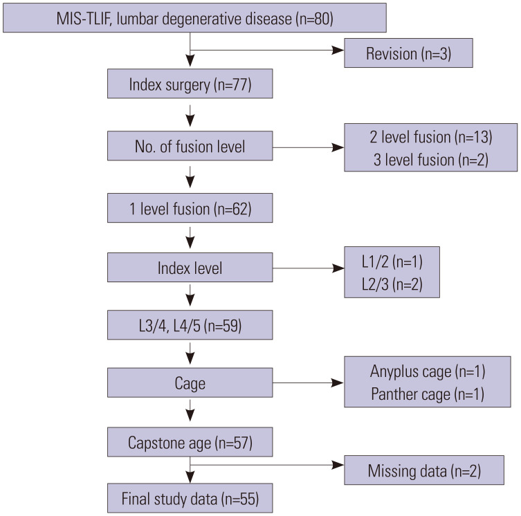
Materials and methods: Fifty-five patients underwent MIS-TLIF at our institute from January 2018 to September 2019. Demographic, pre- and postoperative radiologic, and cage-related factors were included. Statistical analyses were designed to compare patients whose SL increased with decreased after surgery.

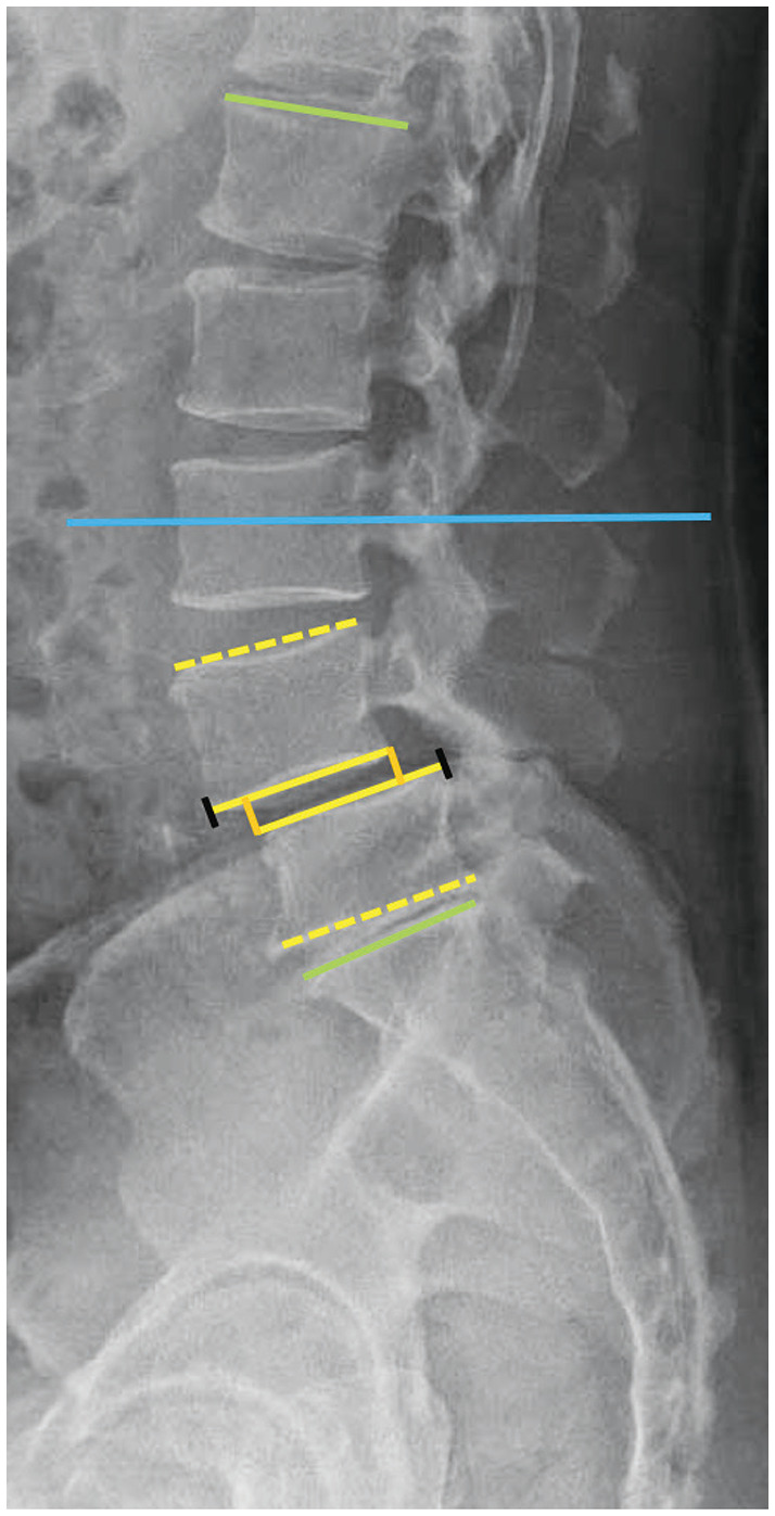
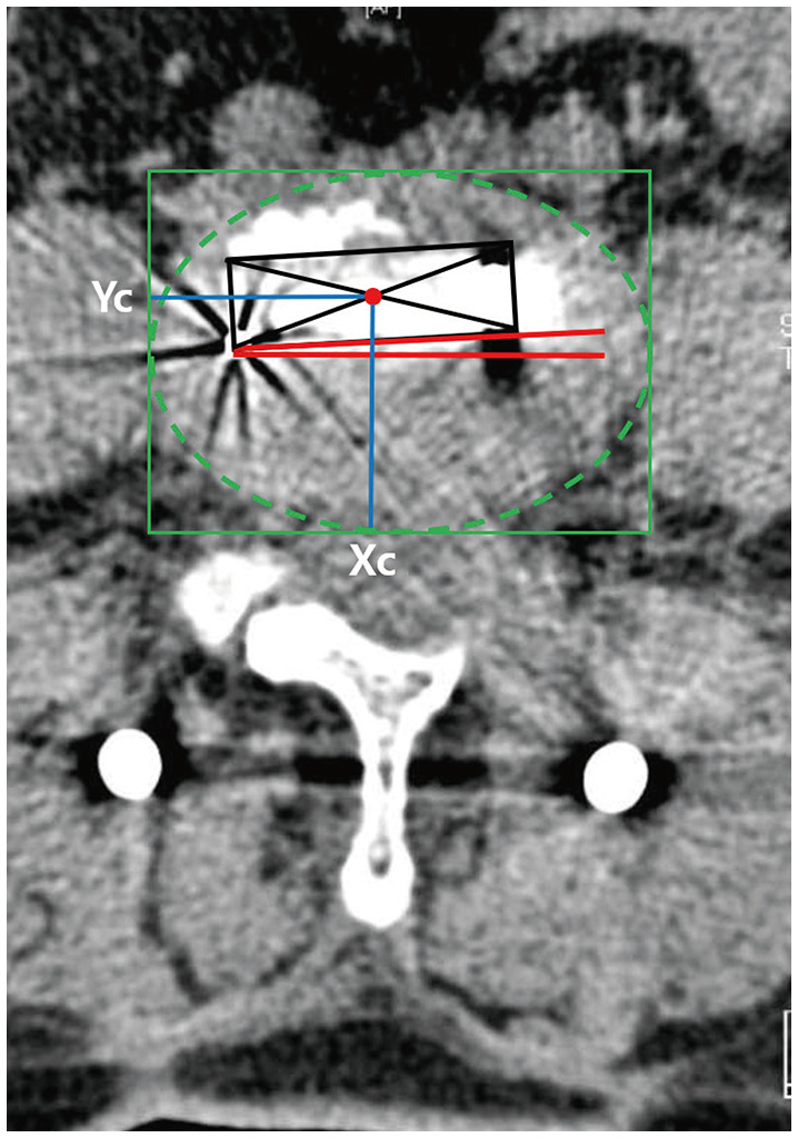
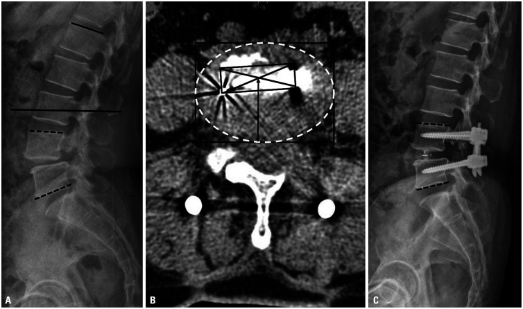
Results: After surgery, SL increased in 34 patients (group I) and decreased in 21 patients (group D). The index level, disc lordosis, SL, lumbar lordosis, proximal lordosis (PL), and Y-axis position of the cage (Yc) differed significantly between groups I and D. The cage in group I was more anterior than that in group D (Yc: 55.84% vs. 51.24%). Multivariate analysis showed that SL decreased more significantly after MIS-TLIF when the index level was L3/4 rather than L4/5 [odds ratio (OR): 0.46, p=0.019], as preoperative SL (OR: 0.82, p=0.037) or PL (OR: 0.68, p=0.028) increased, and as the cage became more posterior (OR: 1.10, p=0.032).

Conclusion: Changes in SL after MIS-TLIF appear to be associated with preoperative SL and PL, index level, and Yc. An index level at L4/5 instead of L3/4, smaller preoperative SL or PL, and an anterior position of the cage are likely to result in increased SL after MIS-TLIF.

Keywords: Minimally invasive; cage; lumbar lordosis; outcome; segmental lordosis; spine surgery; transforaminal lumbar interbody fusion.


Conflict of interest statement

The authors have no potential conflicts of interest to disclose.

Figures

Fig. 1
Fig. 1. Flow diagram for patient selection. MIS-TLIF, minimally invasive transforaminal lumbar interbody fusion.
Fig. 2
Fig. 2. Preoperative radiologic parameters. Disc lordosis (DCL): angle between the inferior endplate of the upper vertebra and the superior endplate of the lower vertebra of the index level (yellow line), Segmental lordosis (SL): angle between the superior endplate of the upper vertebra and the inferior endplate of the lower vertebra of the index level (yellow dotted line). Anterior disc height (DHA): perpendicular distance between the superior endplate of the anterior end of the lower vertebra and the inferior endplate of the upper vertebra of the index level (anterior orange line). Posterior disc height (DHP): perpendicular distance between the inferior endplate at the posterior end of the upper vertebra and the superior endplate of the lower vertebra of the index level (posterior orange line). Lumbar lordosis (LL): angle between the superior endplate of L1 and the superior endplate of S1 (green line). Proximal lordosis (PL): angle between a horizontal line (blue line) and the superior endplate of the L1 vertebra (superior green line). Distal lordosis (DL): angle between a horizontal line and the superior endplate of S1 (inferior green and blue line).
Fig. 3
Fig. 3. Cage location–related radiologic variables. The center of the cage (red circle) was determined using the diagonal intersection of the cage on an axial CT image. The corner of the vertebral body was determined by approximating an ellipse, and the X and Y axes were determined accordingly (green solid line). The cage position (blue line) was converted into a ratio by comparing the X and Y diameters of the ellipse. If Xc was less than 50%, the cage was located on the right, and if it was more than 50%, the cage was located on the left. If Yc was less than 50%, the cage was located at the posterior, and if it was more than 50%, the cage was located at the anterior. The angle of the cage was determined using the long axis of the cage and the X axis of the ellipse (red angle). Therefore, the smaller the angle, the more transversely the cage is positioned. Yc, Y-axis position of the cage; Xc, X-axis position of the cage.
Fig. 4
Fig. 4. Patient with decreased segmental lordosis after surgery even though the cage was positioned anteriorly. This patient had large preoperative segmental lordosis (black dotted line, 26.6°) and proximal lordosis (black line, 17.7°) (A). Although the cage was positioned anteriorly (black arrow, 67.4%) in the axial CT image (B), postoperative segmental lordosis was decreased by 15.8° (black dotted line) on 1-year postoperative X-ray (C).
Fig. 5
Fig. 5. Patient with increased segmental lordosis after surgery even though the cage was not positioned anteriorly. This patient had small preoperative segmental lordosis (black dotted line, 6.2°) and proximal lordosis (black line, 6.7°) (A). Even though the cage was not positioned anteriorly enough (black arrow, 44.4%) in the axial CT image (B), postoperative segmental lordosis increased by 20.5° (black dotted line) on 1-year postoperative X-ray (C).

Similar articles

Cited by

References

    1. Kepler CK, Rihn JA, Radcliff KE, Patel AA, Anderson DG, Vaccaro AR, et al. Restoration of lordosis and disk height after single-level transforaminal lumbar interbody fusion. Orthop Surg. 2012;4:15–20. - PMC - PubMed
    1. Tian H, Wu A, Guo M, Zhang K, Chen C, Li X, et al. Adequate restoration of disc height and segmental lordosis by lumbar interbody fusion decreases adjacent segment degeneration. World Neurosurg. 2018;118:e856–e864. - PubMed
    1. Brodano GB, Martikos K, Lolli F, Gasbarrini A, Cioni A, Bandiera S, et al. Transforaminal lumbar interbody fusion in degenerative disk disease and spondylolisthesis grade I: minimally invasive versus open surgery. J Spinal Disord Tech. 2015;28:E559–E564. - PubMed
    1. Choi WS, Kim JS, Ryu KS, Hur JW, Seong JH. Minimally invasive transforaminal lumbar interbody fusion at L5-S1 through a unilateral approach: technical feasibility and outcomes. Biomed Res Int. 2016;2016:2518394. - PMC - PubMed
    1. Lee WC, Park JY, Kim KH, Kuh SU, Chin DK, Kim KS, et al. Minimally invasive transforaminal lumbar interbody fusion in multilevel: comparison with conventional transforaminal interbody fusion. World Neurosurg. 2016;85:236–243. - PubMed

MeSH terms