. 2022 Sep;55(9):e13273.
doi: 10.1111/cpr.13273. Epub 2022 Jun 24.

DNASE1L3 inhibits proliferation, invasion and metastasis of hepatocellular carcinoma by interacting with β-catenin to promote its ubiquitin degradation pathway

Affiliations

DNASE1L3 inhibits proliferation, invasion and metastasis of hepatocellular carcinoma by interacting with β-catenin to promote its ubiquitin degradation pathway

Bo Li et al. Cell Prolif. 2022 Sep.

Abstract

As a member of the deoxyribonuclease 1 family, DNASE1L3 plays a significant role both inside and outside the cell. However, the role of DNASE1L3 in hepatocellular carcinoma (HCC) and its molecular basis remains to be further investigated. In this study, we report that DNASE1L3 is downregulated in clinical HCC samples and evaluate the relationship between its expression and HCC clinical features. In vivo and in vitro experiments showed that DNASE1L3 negatively regulates the proliferation, invasion and metastasis of HCC cells. Mechanistic studies showed that DNASE1L3 recruits components of the cytoplasmic β-catenin destruction complex (GSK-3β and Axin), promotes the ubiquitination degradation of β-catenin, and inhibits its nuclear transfer, thus, decreasing c-Myc, P21 and P27 level. Ultimately, cell cycle and EMT signals are restrained. In general, this study provides new insight into the mechanism for HCC and suggests that DNASE1L3 can become a considerable target for HCC.


Conflict of interest statement

The authors declare no conflict of interests.

Figures
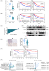
FIGURE 1
FIGURE 1
Expression of DNASE1L3 in hepatocellular carcinoma and its prognostic significance. (A and B) The The Cancer Genome Atlas (TCGA) and Gene Expression Omnibus (GEO) database showed that the transcription level of DNASE1L3 was lower in hepatocellular carcinoma (HCC) compared to paracarcinoma tissues, and high expression of DNASE1L3 has a good overall survival (OS) and progression‐free survival (PFS). (C) The transcription level of DNASE1L3 was detected by RT‐qPCR in 28 pairs of HCC tissues and adjacent non‐tumour tissues. The results showed that 25 (89%) of them were significantly down‐regulated. (D) The expression level of DNASE1L3 protein was detected by Western blot in 12 pairs of HCC tissues and adjacent non‐tumour tissues. (E) The expression level of DNASE1L3 in tissue microarray chip was measured by IHC. The expression level of DNASE1L3 in cancer and paracancerous tissues were compared according to the histochemical score of AI intelligence score. Scale bar: 100 μm. (F) Kaplan–Meier analysis showed that patients with high expression of DNASE1L3 had better OS and DFS. (G) Univariate and multivariate analyses of different clinicopathological features in HCC patients.
FIGURE 2
FIGURE 2
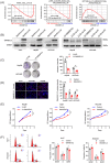
Overexpression of DNASE1L3 inhibits the proliferation of hepatocellular carcinoma cells. Gene enrichment analysis showed that the predefined genomes involved in cell cycle, cell proliferation, cell metastasis and invasion were significantly enriched in low‐level DNASE1L3, indicated that DNASE1L3 may has an inhibitory effect on above properties. (B) Western blot analysis of the expression of DNASE1L3 in hepatocellular carcinoma (HCC) cells transfected with overexpression plasmid or siRNA. (C) Colony formation assays revealed that DNA overexpression inhibited the proliferation of Hep3B and HCCLM3 cells. (D) EdU assay showed that the proliferation of Huh7, HCCLM3 and Hep3B cells decreased after DNASE1L3 overexpression. Scale bar: 200 μm. (E) DNASE1L3 overexpression markedly inhibited cell viability by CCK‐8 assay. (F) Flow cytometry showed that the overexpression of DNASE1L3 increased the G0/G1 phase of Huh7 and HCCLM3 cells and blocked the cell cycle in G1 phase. Student's t‐ test. Mean ± SD (*p < 0.05; **p < 0.01). LV‐D + si‐#1/LV‐D + si‐#2: overexpression of DNASE1L3 by lentivirus, and then transient knockdown of DNASE1L3 using siRNA#1/siRNA#2.
FIGURE 3
FIGURE 3
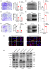
Overexpression of DNASE1L3 inhibits invasion and metastasis of hepatocellular carcinoma cells. (A–C) Transwell assay showed that the metastatic and invasive ability of Huh7, HCCLM3 and Hep3B cells decreased after DNASE1L3 overexpression. Scale bar: 200 μm. (D–F) Wound healing assay showed that the migratory ability of Huh7, HCCLM3 and Hep3B cells decreased after DNASE1L3 overexpression. Scale bar: 200 μm. (G) The relative fluorescence intensities of E‐cadherin and N‐cadherin were assessed by IF staining in Huh7 and HCCLM3 cells after DNASE1L3 overexpression. Scale bar: 20 μm. (H) The expression levels of Vimentin, c‐Myc, N‐cadherin, E‐cadherin, p21 and p27 in HCCLM3 and Hep3B cells were detected by Western blot. Student's t‐test. Mean ± SD (*p < 0.05; **p < 0.01).
FIGURE 4
FIGURE 4
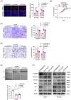
Knockdown of DNASE1L3 promotes proliferation, invasion and metastasis of hepatocellular carcinoma cells. EdU assay showed that the knockdown of DNASE1L3 promoted the proliferation of HCCLM3 and Hep3B cell. Scale bar: 200 μm. (B) CCK‐8 assay revealed that the proliferation of HCCLM3 cell was decreased after DNASE1L3 knockdown, compared with the control. (C and D) Transwell assay demonstrated that knockdown of DNASE1L3 promotes the metastasis and invasion of HCCLM3 and Hep3B cells. Scale bar: 200 μm. (E) Wound healing assay showed that knockdown of DNASE1L3 promotes the migration of Hep3B cells. Scale bar: 200 μm. (F) The expression levels of Vimentin, c‐Myc, N‐cadherin, E‐cadherin, p21 and p27 in Huh7, HCCLM3 and Hep3B cells were detected by Western blot. Student's t‐test. Mean ± SD (*p < 0.05; **p < 0.01).
FIGURE 5
FIGURE 5
DNASE1L3 inhibits tumorigenicity and metastasis of hepatocellular carcinoma in vivo. Dissected tumours from HCCLM3 cells stably expressing DNASE1L3 and control cells. (B) Nude mice were subcutaneously transplanted with HCCLM3 cells stably expressing DNASE1L3 and control cells, and the growth curve and weight of the tumour were obtained. (C) Representative H&E and IHC staining of dissected tumour tissues were shown. Scale bar: 100 μm. (D) Mouse bioluminescence images derived from the IVIS imaging system and representative images of H&E staining of lung tissue. Scale bar: 100 μm. (E) The lung intensity of LV‐NC and LV‐DNASE1L3 groups was quantified by fluorescence intensity. Student's t‐test. Mean ± SD (*p < 0.05; **p < 0.01).
FIGURE 6
FIGURE 6
DNASE1L3 binds to β‐catenin and inhibits its activation. Silver staining of SDS‐PAGE gel. IP was performed by the mixture of Huh7 cell lysis. IgG group was used as control. (B and C) The interaction between DNASE1L3 and β‐catenin in Hep3B cells was determined by exogenous and endogenous Co‐IP and cellular immunofluorescence staining. Scale bar: 20 μm. (D) The mRNA level of β‐catenin was detected by RT‐qPCR after DNASE1L3 overexpression or knockdown. (E) The protein expression of β‐catenin was detected by Western blot after DNASE1L3 overexpression or knockdown. Student's t‐test. Mean ± SD (*p < 0.05; **p < 0.01).
FIGURE 7
FIGURE 7
Correlation between DNASE1L3 and β‐catenin. Pearman correlation of DNASE1L3 and β‐catenin transcription levels were analyzed based on RNA‐seq data from TCGA. (B) Pearson correlation of DNASE1L3 and β‐catenin transcription levels were analyzed based on 28 pairs of hepatocellular carcinoma (HCC) tissues. (C) Pearson correlations of DNASE1L3 and β‐catenin protein levels were analyzed based on 18 pairs of HCC tissues. Student's t‐test. Mean ± SD (*p < 0.05; **p < 0.01).
FIGURE 8
FIGURE 8
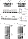
DNASE1L3 promotes β‐catenin ubiquitination and degradation. (A and B) Huh7 and Hep3B cells with or without DNASE1L3 overexpression were treated with MG132 for 8 h, and the fine cytolysis fluid was prepared. The expression of DNASE1L3 and β‐catenin protein was detected by Western blot. (C and D) Western blot showing the effect of DNASE1L3 on β‐catenin stability in Huh7 and Hep3B cells incubated with cycloheximide at different time points. (E and F) Huh7 and Hep3B cells were transfected with ubiquitin plasmid, DNASE1L3 overexpression and control plasmids. After 8 h of MG132 treatment, the cell lysate was treated with anti‐β‐catenin antibody for Co‐IP. Anti‐UB antibody was used to detect Western blot. The total cell lysate was detected by anti‐DNASE1L3, anti‐β‐catenin and anti‐GAPDH.
FIGURE 9
FIGURE 9
DNASE1L3 recruits more β‐catenin destruction complexes. (A) The interaction of DNASE1L3 with β‐catenin, GSK‐3 β and AXIN in Hep3B cells was determined by exogenous Co‐IP. (B) After Hep3B cells were transfected with DNASE1L3 overexpression and control plasmids, Co‐IP was performed with anti‐β‐catenin antibody, and Western blot was detected with anti‐DNASE1L3, anti‐β‐catenin, anti‐GSK‐3β and anti‐AXIN antibodies. (C) The relative fluorescence intensity of β‐catenin and GSK‐3β in DNASE1L3 overexpressed Hep3B cells was measured by exogenous cellular immunofluorescence assay. Scale bar: 20 μm. (D) The protein levels of DNASE1L3 and β‐catenin in cytoplasm and nucleus of DNASE1L3 overexpressed Hep3B cells were detected by the nucleoplasmic separation assay. (E) The cell viability of DNASE1L3‐Ctrl group, DNASE1L3‐OE group, β‐catenin‐OE group and D‐OE + β‐OE group was determined by CCK‐8 and EdU assay. Scale bar: 200 μm. (F) The protein levels of DNASE1L3, N‐cadherin, vimentin, β‐catenin, c‐Myc and GAPDH in DNASE1L3‐Ctrl group, DNASE1L3‐OE group, β‐catenin‐OE group and D‐OE + β‐OE group was detected by Western blot. Student's t‐test. Mean ± SD (*p < 0.05; **p < 0.01). D‐OE + β‐OE: overexpression of DNASE1L3 and β‐catenin by lentivirus.

Similar articles

Cited by

References

    1. Global Burden of Disease Liver Cancer Collaboration , Akinyemiju T, Abera S, et al. The burden of primary liver cancer and underlying etiologies from 1990 to 2015 at the global, regional, and National Level: results from the global burden of disease study 2015. JAMA Oncol. 2017;3(12):1683‐1691. doi:10.1001/jamaoncol.2017.3055 - DOI - PMC - PubMed
    1. Galle PR, Forner A, Llovet JM, et al. EASL Clinical Practice Guidelines: Management of hepatocellular carcinoma. J Hepatol. 2018;69(1):182‐236. doi:10.1016/j.jhep.2018.03.019 - DOI - PubMed
    1. Forner A, Reig M, Bruix J. Hepatocellular carcinoma. Lancet. 2018;391(10127):1301‐1314. doi:10.1016/S0140-6736(18)30010-2 - DOI - PubMed
    1. Jiang X, Wang G, Liu Y, et al. A novel long non‐coding RNA RP11‐286H15.1 represses hepatocellular carcinoma progression by promoting ubiquitination of PABPC4. Cancer Lett. 2021;499:109‐121. doi:10.1016/j.canlet.2020.11.038 - DOI - PubMed
    1. Zhong JH, Ke Y, Gong WF, et al. Hepatic resection associated with good survival for selected patients with intermediate and advanced‐stage hepatocellular carcinoma. Ann Surg. 2014;260(2):329‐340. doi:10.1097/SLA.0000000000000236 - DOI - PubMed

MeSH terms