

Advances in the ultrasound diagnosis in equine reproductive medicine: New approaches
- PMID: 35748405
- PMCID: PMC9796632
- DOI: 10.1111/rda.14192
Advances in the ultrasound diagnosis in equine reproductive medicine: New approaches
Abstract
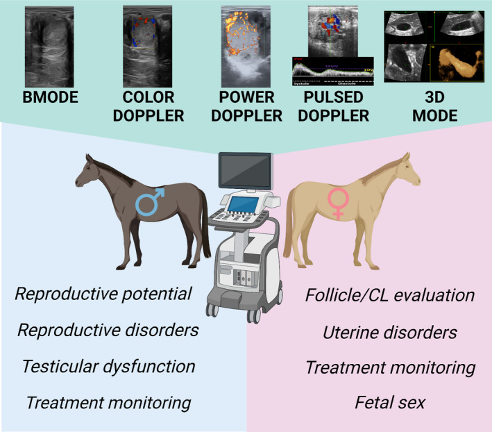
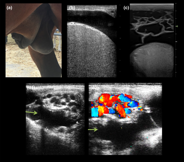
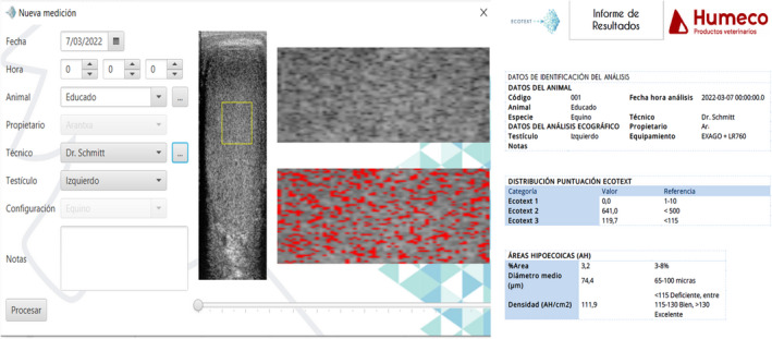
Ultrasound technology has led to new lines of research in equine reproduction, and it has helped to greatly improve clinical diagnosis and reproductive outcomes in equine practice. This review aims to discuss the potential clinical uses and new approaches of ultrasonography in equine reproduction. Doppler modalities are usually used to evaluate the vascularization of the follicles, corpus luteum (CL), and the uterus in the mare for diagnostic purposes. Inclusion of Doppler ultrasound in artificial insemination and embryo transfer programs could improve the reproductive outcome of these techniques. Better selection of recipients based on CL functionality, early pregnancy diagnosis 7-8 days postovulation of the donor before flushing or diagnosis of mares with endometritis with pathological increases of blood flow are examples of clinical applications in the mare. In the stallion, colour Doppler ultrasound has improved the diagnostic potential of B-mode ultrasound, improving the differential diagnosis of pathologies such as testicular torsion (decrease or absence of blood flow in the cord) and orchitis (increased blood flow in the cord). The incorporation of pulsed Doppler ultrasound into the reproductive evaluation of the stallion has enabled early identification of stallions with testicular dysfunction, thus allowing administration of timely treatment and subsequent improvements of the fertility prognosis for these animals. In addition, this technique has been used in the monitoring of patients undergoing medical and surgical treatments, thus verifying their efficacy. Recently, computer-assisted pixel analysis using specific software has been performed in research work in order to semi-quantitatively evaluate the vascularization (colour and power Doppler) and echotexture of different organs. These softwares are now being developed for clinical purposes, as is the case with Ecotext, a computer program developed for the evaluation of testicular echotexture, providing information on testicular functionality.
Keywords: 3D ultrasound; grey scale image; mare; power and colour Doppler; pregnancy diagnosis; pulse Doppler; stallion; subfertility.
© 2022 The Authors. Reproduction in Domestic Animals published by Wiley-VCH GmbH.
Conflict of interest statement
None of the authors have any conflict of interest to declare.
Figures
References
-
- Abdelnaby, E. A. , Emam, I. A. , Salem, N. Y. , Ramadan, E. S. , Khattab, M. S. , Farghali, H. A. , & Abd El Kader, N. A. (2020). Uterine hemodynamic patterns, oxidative stress, and chromoendoscopy in mares with endometritis. Theriogenology, 158, 112–120. 10.1016/j.theriogenology.2020.09.012 - DOI - PubMed
-
- Abecia, J. A. , Carvajal‐Serna, M. , Casao, A. , Palacios, C. , Pulinas, L. , Keller, M. , Chemineau, P. , & Delgadillo, J. A. (2020). The continuous presence of ewes in estrus in spring influences testicular volume, testicular echogenicity and testosterone concentration, but not LH pulsatility in rams. Animal, 14(12), 2554–2561. 10.1017/S1751731120001330 - DOI - PubMed
-
- Acosta, T. J. , Gastal, E. L. , Gastal, M. O. , Beg, M. A. , & Ginther, O. J. (2004). Differential blood flow changes between the future dominant and subordinate follicles precede diameter changes during follicle selection in Mares1. Biology of Reproduction, 71(2), 502–507. 10.1095/biolreprod.104.027896 - DOI - PubMed
-
- Ahmadi, B. , Mirshahi, A. , Giffin, J. , Oliveira, M. E. F. , Gao, L. , Hahnel, A. , & Bartlewski, P. M. (2013). Preliminary assessment of the quantitative relationships between testicular tissue composition and ultrasonographic image attributes in the ram. The Veterinary Journal, 198(1), 282–285. 10.1016/j.tvjl.2013.06.001 - DOI - PubMed
-
- Altermatt, J. L. , Marolf, A. J. , Wrigley, R. H. , & Carnevale, E. M. (2012). Effects of FSH and LH on ovarian and follicular blood flow, follicular growth and oocyte developmental competence in young and old mares. Animal Reproduction Science, 133(3–4), 191–197. 10.1016/j.anireprosci.2012.06.021 - DOI - PubMed
Publication types
MeSH terms
LinkOut - more resources
Full Text Sources
