. 2022 Jun 24;8(25):eabm6756.
doi: 10.1126/sciadv.abm6756. Epub 2022 Jun 24.

Human organ rejuvenation by VEGF-A: Lessons from the skin

Affiliations

Human organ rejuvenation by VEGF-A: Lessons from the skin

Aviad Keren et al. Sci Adv. .

Abstract

Transplanting aged human skin onto young SCID/beige mice morphologically rejuvenates the xenotransplants. This is accompanied by angiogenesis, epidermal repigmentation, and substantial improvements in key aging-associated biomarkers, including ß-galactosidase, p16ink4a, SIRT1, PGC1α, collagen 17A, and MMP1. Angiogenesis- and hypoxia-related pathways, namely, vascular endothelial growth factor A (VEGF-A) and HIF1A, are most up-regulated in rejuvenated human skin. This rejuvenation cascade, which can be prevented by VEGF-A-neutralizing antibodies, appears to be initiated by murine VEGF-A, which then up-regulates VEGF-A expression/secretion within aged human skin. While intradermally injected VEGF-loaded nanoparticles suffice to induce a molecular rejuvenation signature in aged human skin on old mice, VEGF-A treatment improves key aging parameters also in isolated, organ-cultured aged human skin, i.e., in the absence of functional skin vasculature, neural, or murine host inputs. This identifies VEGF-A as the first pharmacologically pliable master pathway for human organ rejuvenation in vivo and demonstrates the potential of our humanized mouse model for clinically relevant aging research.


Figures

Fig. 1.
Fig. 1.. Overview: experimental design.
(A) Aged skin was transplanted onto young (OiY) or old (OiO) mice. Similarly, young skin was transplanted on to young (YiY) or old (YiO) mice. (B) Each group of mice was separated to three groups treated with anti–VEGF-A, VEGF-A, or BSA. Qualitative and quantitative (immuno-)histomorphometry were performed before and 1, 2, and 4 weeks after transplantation. (C) Experimental manipulations in aged human skin transplanted onto young SCID mice (OiY) for 1 or 12 months, along with selected changes in key aging readouts. OiY mice were treated with anti–VEGF-A antibodies, while control groups and OiO mice received intradermal injection of VEGF-A–loaded nanoparticles. (D) Organ culture of aged human skin or epidermal sheets, with/without VEGF-A added to the serum-free culture medium.
Fig. 2.
Fig. 2.. Epidermal and dermal parameters of human aged skin before and after transplantation onto old and young mice.
Before transplantation: Rete-ridge structures were clearly observed in sections of human young skin, whereas old skin is characterized by a marked flattening of the dermoepidermal junction. Increased number of blood vessels and organized collagen in the dermis of the young skin in contrast to the aged one. After transplantation: (A) Increased epidermal thickness (N = 4 young donors, 4 old donors, 5 OiY mice, and 5 OiO mice), (B) Proliferation (N = 4 old donors, 8 OiO mice, and 6 OiY mice) and (C) melanocytes (N = 4 old donors, 9 OiO mice, and 8 OiY mice) in OiY mice compared with pretransplanted aged skin and OiO transplants. (D) p16ink4a expression (N = 3 old donors, 6 OiO mice, and 6 OiY mice) in aged skin before transplantation in OiO and OiY mice and the absence in OiY mice. (E) PGC1α expression in aged skin before transplantation in OiO and OiY (N = 4 old donors, 8 OiO mice, and 6 OiY mice). (F) Expression of SIRT1 (N = 4 old donors, 7 OiO mice, and 6 OiY mice). (G) MTCO-1 (N = 4 old donors, 7 OiO mice, and 7 OiY mice). (H) Quantitation. Data were assessed by IHC from four individual donors. Four areas were evaluated per section, and three sections were analyzed per mouse. After the Shapiro-Wilk test, unpaired Student’s t test: *P < 0.05, **P < 0.01, and ***P < 0.001 or nonparametric Mann-Whitney U test: ###P < 0.001. EP, epidermis; DER, dermis; SC, stratum corneum; SG, stratum granulosum; SS, startum spinosum; SB, stratum basale; PL, papillary layer; RL, reticular layer; H&E, hematoxylin and eosin; N.S., not significant. Scale bars, 50 μm.
Fig. 3.
Fig. 3.. Biomarkers related to epidermal skin aging.
(A) The absence of epidermal filaggrin (N = 4 old donors, 8 OiO mice, and 8 OiY mice) in aged skin before transplantation and in OiO mice versus reappearance in YiO mice. (B) COL17A1 expression (N = 3 old donors, 7 OiO mice, and 7 OiY mice) along the basement membrane of pre-engrafted skin and in the OiO compared to the OiY xenotransplants and (C) MMP1 (N = 3 old donors, 7 OiO mice, and 6 OiY mice). (D) A structural disorganization and decrease in collagen fibers in the pretransplanted aged skin just as in OiO mice, while complete recovery along the dermis of OiY mice (N = 4 old donors, 7 OiO mice, and 6 OiY mice). (E) Quantitation. Data were assessed by IHC from three individual donors. Four areas were evaluated per section, and three sections were analyzed per mouse. After the Shapiro-Wilk test, unpaired Student’s t test: *P < 0.05, and **P < 0.01. EP, epidermis; DER, dermis; SS, startum spinosum. Scale bars, 50 μm.
Fig. 4.
Fig. 4.. Density of human dermal blood vessels before and after transplantation onto old and young mice.
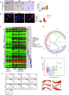
(A) A significantly increased number of CD31+ blood vessels in OiY mice compared with OiO and pretransplanted skin. (B) Double staining of human (red; arrows) and murine (green; arrowheads). EP, epidermis; DER, dermis. Scale bars, 50 μm. (C) RNA-seq analyses in OiY mice. DEGs in the first week versus pretransplant included numerous transcripts related to angiogenesis and for 2 and 4 weeks after transplantation. (D) SCTST on the RNA-seq data identified six trajectory clusters. (E) Volcano plots of the distribution of –log10 (P values) versus the gene expression fold changes. Genes with fold change >2 and P value <0.05 are indicated in green, and genes with fold change <−2 and P value <0.05 are indicated in red. The up-regulated DEGs related to promoting angiogenesis are overimposed on the volcano plot, as well as the down-regulated DEGs that are known to inhibit angiogenesis. (F) qRT-PCR time series profiles of the up-regulation of HIF1A, as well as angiogenesis-related genes such as CXCL1 and CXCL5, MMP9, PGF, LCN2, and ESM1. The red and blue lines mark the expression trajectories of two different patients.
Fig. 5.
Fig. 5.. Biomarkers related to epidermis of aged skin in pretransplanted skin in YiO and after 12 months on the mice.
Expression of (A) MMP1 (N = 3 old donors, 4 OiY mice after 1 month, and 6 OiY mice after 12 months), (B) PGC1α (N = 3 old donors, 5 OiY mice after 1 month, and 6 OiY mice after 12 months), (C) SIRT1 (N = 3 old donors, 5 OiY mice after one month, and 6 OiY mice after 12 months), and (D) MTCO-1 (N = 3 old donors, 4 OiY mice after 1 month, and 6 OiY mice after 12 months) in epidermis of pretransplanted aged skin, in OiY and in 12-month-aged transplants. (E) Quantitation. Four areas were evaluated per section, and three sections were analyzed per mouse. After the Shapiro-Wilk test, Student’s t test: *P < 0.05. EP, epidermis; DER, dermis. Scale bars, 50 μm.
Fig. 6.
Fig. 6.. VEGF-A RNA and protein in OiO and OiY xenotransplants.
(A) In situ hybridization for human VEGF-A in OiO and OiY transplants (N = 3 OiY mice and 3 OiO mice). (B) Quantification of the number of hVEGF-A+ cells in n = 3 xenotransplants per group. Means ± SEM, n = 9 microscopic fields per xenotransplant. For each xenotransplant, n = 3 nonconsecutive sections were evaluated, and for each section, n = 3 microscopic fields were analyzed. (C and D) VEGF-A protein expression in OiO (N = 4 mice) versus OiY (N = 3 mice). (E) Quantitative analysis. Data were assessed by IHC from three individual donors. Four areas were evaluated per section, and three sections were analyzed per mouse. After the Shapiro-Wilk test, Student’s t test or nonparametric Mann-Whitney test for ISH: *P < 0.05, and **P < 0.01. Scale bars, 50 μm.
Fig. 7.
Fig. 7.. Expression of VEGF-A by keratinocytes, macrophages, and platelets in pretransplanted aged skin, in OiO, and in OiY xenotransplants.
Double IHC staining revealed decreased VEGF-A protein expression (A) by cytokeratin 10 (expressed by keratinocytes) (N = 3 old donors, 5 OiO mice, and 7 OiY mice), (B) by CD68 (macrophages) (N = 3 old donors, 5 OiO mice, and 7 OiY mice), and by (C) CD42b (platelets) (N = 4 old donors, 5 OiO mice, and 5 OiY mice). (D) Quantitative analysis. Data were assessed by IHC from three individual donors. Four areas were evaluated per section, and three sections were analyzed per mouse. Nonparametric Mann-Whitney U test: ##P < 0.01 and ###P < 0.001. Scale bars, 50 μm.
Fig. 8.
Fig. 8.. Epidermal and dermal parameters of human aged skin before and after transplantation onto young mice treated with VEGF-blocking antibodies.
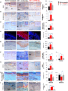
Each group included 11 OiY mice transplanted with skin from six human donors (age range, 77 to 83 years; mean, 81). Injections to OiY decreased skin aging–related biomarkers compared to OiY injected with isotype control as follows: (A) epidermal thickness, (B) proliferation (Ki-67+ keratinocytes), (C) number of melanocytes (Melan-A+ cells), (D) PGC1α, (E) SIRT1, (F) mast cells (c-KIT+), and (G) CD31+ blood vessels, as well as the (H) proportion of thick and thin collagen fibers and (I) collagen 17A. (J) Quantitation. For all tested markers: N = 4 old donors, 6 OiY mice injected with control Abs, and 8 OiY mice injected with VEGF-blocking Abs. Data were assessed by IHC from four individual donors. Four areas were evaluated per section, and three sections were analyzed per mouse. After the Shapiro-Wilk test, Student’s t test: *P < 0.05, **P < 0.01, and ***P < 0.001, or nonparametric Mann-Whitney U test: ##P < 0.01. EP, epidermis; DER, dermis; SG, stratum granulosum; SB, stratum basale; PL, papillary layer; RL, reticular layer; Abs, antibodies. Scale bars, 50 μm.
Fig. 9.
Fig. 9.. Epidermal and dermal parameters of human aged skin before and after transplantation onto young mice treated with VEGF-A loaded nanoparticles.
Intradermal injections to OiO mice increased skin aging–related biomarkers compared to OiO injected with BSA-loaded PLGA nanoparticles and to aged skin before transplantation. The biomarkers were as follows: (A) epidermal thickness (N = 3 old donors, 7 OiO mice injected with PLGA-VEGF, and 6 OiO mice injected with PLGA-BSA), (B) proliferation (Ki-67+ keratinocytes) (N = 3 old donors, 7 OiO mice injected with PLGA-VEGF, and 6 OiO mice injected with PLGA-BSA), and (C) differentiation (filaggrin) (N = 3 old donors, 7 OiO mice injected with PLGA-VEGF, and 7 OiO mice injected with PLGA-BSA), (D) number of melanocytes (Melan-A+ cells) (N = 3 old donors, 8 OiO mice injected with PLGA-VEGF, and 6 OiO mice injected with PLGA-BSA), (E) mast cells (c-Kit+) (N = 3 old donors, 8 OiO mice injected with PLGA-VEGF, and 7 OiO mice injected with PLGA-BSA), (F) CD31+ blood vessels (N = 3 old donors, 7 OiO mice injected with PLGA-VEGF, and 6 OiO mice injected with PLGA-BSA), and (G) increased thick dermal collagen bundles while thin collagen filaments decreased and quantitation (N = 3 old donors, 7 OiO mice injected with PLGA-VEGF, and 7 OiO mice injected with PLGA-BSA). (H) Quantitation. Data were assessed by IHC from four individual donors. Four areas were evaluated per section, and three sections were analyzed per mouse. After the Shapiro-Wilk test, Student’s t test: *P < 0.05 and **P < 0.01, or nonparametric Mann-Whitney U test: #P < 0.05. EP, epidermis; DER, dermis; SG, stratum granulosum; SB, stratum basale; PL, papillary layer; RL, reticular layer. Scale bars, 50 μm.
Fig. 10.
Fig. 10.. Schematic representation of human aged skin before and after transplantation onto young SCID beige mice.
(A) Schematic illustration showing the characteristic changes seen in human skin aging process with decreased epidermal thickness and proliferation, flattened epidermal rete ridges, senescent dermal fibroblasts, epidermal keratinocytes and melanocytes, impaired angiogenesis, decreased upstream and downstream regulators involved in the induction of VEGF-A, and increased biomarker signatures of skin aging. (B) Summary of the proposed rejuvenation cascade of aged human skin after transplantation to young mice (OiY). High-level mouse VEGF-A may initiate this cascade by stimulating human VEGFRs in the xenotransplant, leading to a positive feedback loop that enhances the secretion of human VEGF-A within in the xenotransplant and triggers the indicated cascade of proangiogenic and skin rejuvenation signaling events. The rejuvenation effect can be antagonized significantly, although not entirely abrogated, by neutralizing antibodies that recognize both human and mouse VEGF-A. Thus, additional signals besides VEGF-A secretion that emanate from young mice may contribute to the up-regulation of VEGF-A within the human, namely, mouse-derived signals that up-regulate HIFα, PGC1α, LCN2, and FUT2 expression in human skin. Abs, antibodies; MMP1, matrix metallopeptidase 1; ROS, reactive oxygen species; SEMA3A, semaphorin 3A; SASP, senescence-associated secretory phenotype; RR, rete ridges.

References

    1. D. A. Sinclair, M. D. LaPlante, Lifespan: Why We Age—And Why We Don’t Have To (Atria Books, 2019).
    1. Mkrtchyan G. V., Abdelmohsen K. P., Andreux P., Bagdonaite I., Barzilai N., Brunak S., Cabreiro F., de Cabo R., Campisi J., Cuervo A. M., Demaria M., Ewald C. Y., Fang E. F., Faragher R., Ferrucci L., Freund A., Silva-García C. G., Georgievskaya A., Gladyshev V. N., Glass D. J., Gorbunova V., de Grey A., He W.-W., Hoeijmakers J., Hoffmann E., Horvath S., Houtkooper R. H., Jensen M. K., Jensen M. B., Kane A., Kassem M., de Keizer P., Kennedy B., Karsenty G., Lamming D. W., Lee K.-F., Aulay N. M., Mamoshina P., Mellon J., Molenaars M., Moskalev A., Mund A., Niedernhofer L., Osborne B., Pak H. H., Parkhitko A., Raimundo N., Rando T. A., Rasmussen L. J., Reis C., Riedel C. G., Franco-Romero A., Schumacher B., Sinclair D. A., Suh Y., Taub P. R., Toiber D., Treebak J. T., Valenzano D. R., Verdin E., Vijg J., Young S., Zhang L., Bakula D., Zhavoronkov A., Scheibye-Knudsen M., ARDD 2020: From aging mechanisms to interventions. Aging (Albany NY) 12, 24484–24503 (2020). - PMC - PubMed
    1. Kirkland J. L., Tchkonia T., Zhu Y., Niedernhofer L. J., Robbins P. D., The clinical potential of senolytic drugs. J. Am. Geriatr. Soc. 65, 2297–2301 (2017). - PMC - PubMed
    1. Schumacher B., Krieg T. M., The aging skin: From basic mechanisms to clinical applications. J. Invest. Dermatol. 141, 949–950 (2021). - PubMed
    1. Stone R. C., Aviv A., Paus R., Telomere dynamics and telomerase in the biology of hair follicles and their stem cells as a model for aging research. J. Invest. Dermatol. 141, 1031–1040 (2021). - PubMed

LinkOut - more resources