

Histomorphological features of mucormycosis with rise and fall of COVID-19 pandemic
- PMID: 35749915
- PMCID: PMC9212773
- DOI: 10.1016/j.prp.2022.153981
Histomorphological features of mucormycosis with rise and fall of COVID-19 pandemic
Abstract
Introduction: Due to Corona Virus disease -19, India saw a surge of mucormycosis cases, associated with high death rate. India, during the month of May to July 2021 saw a surge of mucormycosis from all states, with close to 50,000 cases just in a span of 3 months.
Objective: To examine the histopathological appearances of rhino-orbital/rhino-maxillary/sino-nasal mucormycosis in the backdrop of the ongoing COVID 19 pandemic.
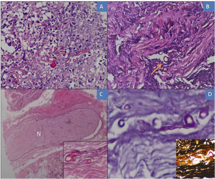
Material and methods: The study involved analysis of 60 biopsy samples of suspected rhino-maxillary /rhino-orbital mucormycosis received from post-COVID-19 patients. A preliminary review of the slides showing hyphal forms of fungal organisms with un-doubtful tissue / mucosal invasion was included. All samples were examined under Hematoxylin and Eosin stains along with special fungal stains. Data thus obtained were analyzed statistically. Special stains for fungus namely Periodic Acidic Schiff (PAS) and Gomori Methenamine silver (GMS) were utilized to confirm and/or to differentiate the fungal organisms and to highlight the cell wall of the fungus.
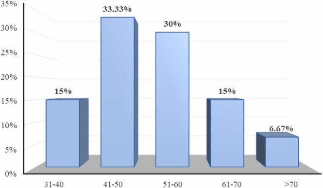
Results: The mean age of the patients with mucormycosis was 51.68 years and 72 (83.33%) of them were males. Acute type of inflammation was noted in 44 (73.33%), granulomatous inflammation in 14 (23.33%) of cases. Bony invasion and perineural invasion was observed in 5 (8.33%) and 55 (91.67%) cases, respectively. The dominant fungus were mucorales in 58 (96.67%), aspergillous, along with mucorales in 12 (20%) and combination of mucorales and candida identified in 8 (13.33%) cases.
Conclusion: Besides all the histological appearance of angioinvasion, bone, and soft tissue invasion, a notable aspect was the shift in inflammatory pattern, which was more granulomatous in nature, with a decrease in fungal load correlating with the drop of COVID second wave. This proves that as immunity develops, the host's response to secondary opportunistic infections changes.
Keywords: COVID-19; Mucorales; Mucormycosis / microbiology; Mycoses / complications; SARS-CoV-2.
Copyright © 2022 Elsevier GmbH. All rights reserved.
Conflict of interest statement
Authors have declared that no financial / conflict of interest to disclose.
Figures


Similar articles
-
Rhinocerebral mucormycosis: Pathology revisited with emphasis on perineural spread.Neurol India. 2014 Jul-Aug;62(4):383-6. doi: 10.4103/0028-3886.141252. Neurol India. 2014. PMID: 25237943
-
Spectrum of orbital fat necrosis in rhino-orbital-cerebral mucormycosis in post-COVID-19 patients.Indian J Ophthalmol. 2024 Oct 1;72(10):1478-1482. doi: 10.4103/IJO.IJO_2623_23. Epub 2024 Sep 27. Indian J Ophthalmol. 2024. PMID: 39331438 Free PMC article.
-
Role of histopathology in severity assessments of post-COVID-19 rhino-orbital cerebral mucormycosis - A case-control study.Ann Diagn Pathol. 2023 Dec;67:152183. doi: 10.1016/j.anndiagpath.2023.152183. Epub 2023 Jul 27. Ann Diagn Pathol. 2023. PMID: 37696132
-
Immunopathology of COVID-19 and its implications in the development of rhino-orbital-cerebral mucormycosis: a major review.Orbit. 2022 Dec;41(6):670-679. doi: 10.1080/01676830.2022.2099428. Epub 2022 Jul 20. Orbit. 2022. PMID: 35856238 Review.
-
Surge of mucormycosis during the COVID-19 pandemic.Travel Med Infect Dis. 2023 Mar-Apr;52:102557. doi: 10.1016/j.tmaid.2023.102557. Epub 2023 Feb 20. Travel Med Infect Dis. 2023. PMID: 36805033 Free PMC article. Review.
Cited by
-
An old confusion: Entomophthoromycosis versus mucormycosis and their main differences.Front Microbiol. 2022 Nov 3;13:1035100. doi: 10.3389/fmicb.2022.1035100. eCollection 2022. Front Microbiol. 2022. PMID: 36406416 Free PMC article. Review.
-
Acute invasive fungal rhinosinusitis (AIFRS) - A histopathological analysis of expanding spectrum of fungal infections in backdrop of COVID-19 pandemic.J Family Med Prim Care. 2023 Sep;12(9):2097-2102. doi: 10.4103/jfmpc.jfmpc_629_23. Epub 2023 Sep 30. J Family Med Prim Care. 2023. PMID: 38024940 Free PMC article.
-
Unexpected Discrepancies in the Histopathological and Microbiological Diagnoses of Suspected Mucormycosis Cases at a South Indian Tertiary Care Center During the COVID-19 Pandemic.Cureus. 2024 Dec 10;16(12):e75478. doi: 10.7759/cureus.75478. eCollection 2024 Dec. Cureus. 2024. PMID: 39791083 Free PMC article.
-
Successful Primary Oral Isavuconazole Therapy in Acute Invasive Fungal Sinusitis with Triple Fungal Species and Multiple Comorbidities.Indian J Otolaryngol Head Neck Surg. 2025 Feb;77(2):1033-1036. doi: 10.1007/s12070-024-05247-w. Epub 2024 Dec 3. Indian J Otolaryngol Head Neck Surg. 2025. PMID: 40070726
-
Invasive Fungal Rhinosinusitis: The First Histopathological Study in Vietnam.Head Neck Pathol. 2024 Oct 16;18(1):104. doi: 10.1007/s12105-024-01711-9. Head Neck Pathol. 2024. PMID: 39412604
References
MeSH terms
LinkOut - more resources
Full Text Sources
Medical
Miscellaneous
