

Phagocytic microglia and macrophages in brain injury and repair
- PMID: 35751629
- PMCID: PMC9344092
- DOI: 10.1111/cns.13899
Phagocytic microglia and macrophages in brain injury and repair
Abstract
Aims: Phagocytosis is the cellular digestion of extracellular particles, such as pathogens and dying cells, and is a key element in the evolution of central nervous system (CNS) disorders. Microglia and macrophages are the professional phagocytes of the CNS. By clearing toxic cellular debris and reshaping the extracellular matrix, microglia/macrophages help pilot the brain repair and functional recovery process. However, CNS resident and invading immune cells can also magnify tissue damage by igniting runaway inflammation and phagocytosing stressed-but viable-neurons.
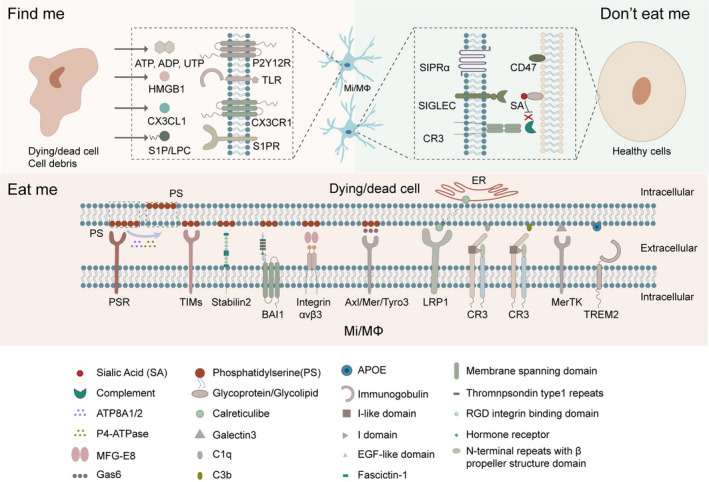
Discussion: Microglia/macrophages help mediate intercellular communication and react quickly to the "find-me" signals expressed by dead/dying neurons. The activated microglia/macrophages then migrate to the injury site to initiate the phagocytic process upon encountering "eat-me" signals on the surfaces of endangered cells. Thus, healthy cells attempt to avoid inappropriate engulfment by expressing "do not-eat-me" signals. Microglia/macrophages also have the capacity to phagocytose immune cells that invade the injured brain (e.g., neutrophils) and to regulate their pro-inflammatory properties. During brain recovery, microglia/macrophages engulf myelin debris, initiate synaptogenesis and neurogenesis, and sculpt a favorable extracellular matrix to support network rewiring, among other favorable roles. Here, we review the multilayered nature of phagocytotic microglia/macrophages, including the molecular and cellular mechanisms that govern microglia/macrophage-induced phagocytosis in acute brain injury, and discuss strategies that tap into the therapeutic potential of this engulfment process.
Conclusion: Identification of biological targets that can temper neuroinflammation after brain injury without hindering the essential phagocytic functions of microglia/macrophages will expedite better medical management of the stroke recovery stage.
Keywords: acute brain injury; brain repair; microglia/macrophage; phagocytosis.
© 2022 The Authors. CNS Neuroscience & Therapeutics published by John Wiley & Sons Ltd.
Conflict of interest statement
None.
Figures
References
-
- Deshpande OA, Wadhwa R. Phagocytosis. StatPearls; 2021. - PubMed
Publication types
MeSH terms
Grants and funding
LinkOut - more resources
Full Text Sources
Miscellaneous
