. 2022 Dec;32(12):8296-8305.
doi: 10.1007/s00330-022-08930-0. Epub 2022 Jun 25.

Development and validation of an ultrasound-based prediction model for differentiating between malignant and benign solid pancreatic lesions

Affiliations

Development and validation of an ultrasound-based prediction model for differentiating between malignant and benign solid pancreatic lesions

Jiayan Huang et al. Eur Radiol. 2022 Dec.

Abstract

Objective: To identify the diagnostic ability of precontrast and contrast-enhanced ultrasound (CEUS) in differentiating between malignant and benign solid pancreatic lesions (MSPLs and BSPLs) and to develop an easy-to-use diagnostic nomogram.

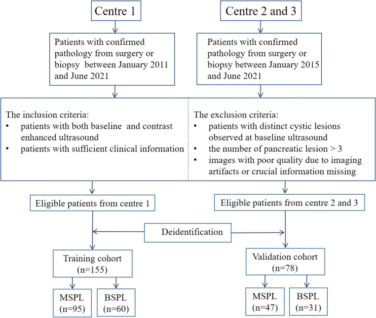
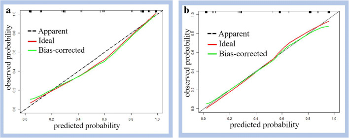
Materials and methods: This study was approved by the institutional review board. Patients with pathologically confirmed solid pancreatic lesions were enrolled from one tertiary medical centre from March 2011 to June 2021 and in two tertiary institutions between January 2015 and June 2021. A prediction nomogram model was established in the training set by using precontrast US and CEUS imaging features that were independently associated with MSPLs. The performance of the prediction model was further externally validated.

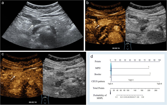
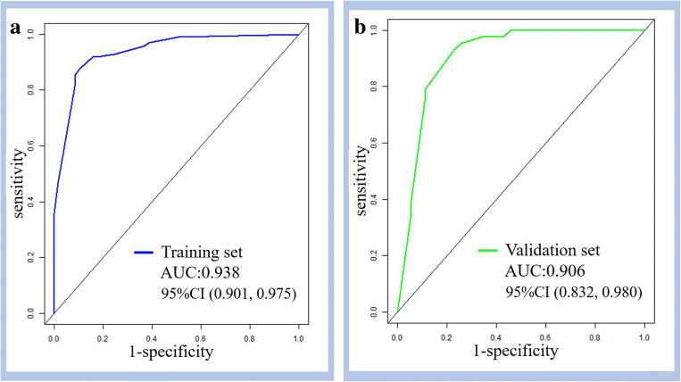
Results: A total of 155 patients (mean age, 55 ± 14.6 years, M/F = 84/71) and 78 patients (mean age, 59 ± 13.4 years, M/F = 36/42) were included in the training and validation cohorts, respectively. In the training set, an ill-defined border and dilated main pancreatic duct on precontrast ultrasound, CEUS patterns of hypoenhancement in both the arterial and venous phases of CEUS, and hyperenhancement/isoenhancement followed by washout were independently associated with MSPLs. The prediction nomogram model developed with the aforementioned variables showed good performance in differentiating MSPLs from BSPLs with an area under the curve (AUC) of 0.938 in the training set and 0.906 in the validation set.

Conclusion: Hypoenhancement in all phases, hyperenhancement/isoenhancement followed by washout on CEUS, an ill-defined border, and a dilated main pancreatic duct were independent risk factors for MSPLs. The nomogram constructed based on these predictors can be used to diagnose MSPLs.

Key points: • An ill-defined border and dilated main pancreatic duct on precontrast ultrasound, hypoenhancement in all phases of CEUS, and hyperenhancement/isoenhancement followed by washout were independently associated with MSPLs. • The ultrasound-based prediction model showed good performance in differentiating MSPLs from BSPLs with an AUC of 0.938 in the training set and 0.906 in the external validation set. • An ultrasound-based nomogram is an easy-to-use tool to differentiate between MSPLs and BSPLs with high efficacy.

Keywords: Contrast media; Nomograms; Pancreatic neoplasms; Ultrasonography.


Conflict of interest statement

The authors of this manuscript declare no relationships with any companies, whose products or services may be related to the subject matter of the article.

Figures

Fig. 1
Fig. 1
Patient selection flowchart. MSPL, malignant solid pancreatic lesion, BSPL, benign solid pancreatic lesion
Fig. 2
Fig. 2
Contrast-enhanced US of a 51-year-old woman with a 2.0-cm hypoechoic solid lesion (arrow) in the neck of the pancreas. a The lesion (arrow) had a clear border, and the diameter of the main pancreatic duct was within the normal range. b Arterial phase hyperenhancement (arrow) was seen on contrast-enhanced US. c The lesion showed slight hyperenhancement (arrow) relative to the surrounding pancreatic parenchyma in the venous phase. d The lesion received a score of 0 according to the nomogram, corresponding to a less than 10% probability of malignancy. A neuroendocrine tumour with pathological differentiation of grade 1 was confirmed by pathological analysis
Fig. 3
Fig. 3
Contrast-enhanced US of a 60-year-old woman with a 5.3-cm hypoechoic solid lesion in the head of the pancreas. a The lesion showed an ill-defined border (arrow) and dilated main pancreatic duct measuring 9 mm. b, c Hypoenhancement of the tumour was demonstrated in both the arterial phase (b) and venous phases (c). d A total of 260 points were assigned to the lesion according to the nomogram, corresponding to a higher than 90% probability of being a malignant solid pancreatic lesion. Poorly differentiated pancreatic ductal adenocarcinoma was confirmed by histopathology
Fig. 4
Fig. 4
The predictive reliability and goodness of fit of the logistic regression were assessed by using the Hosmer–Lemeshow test, showing p values of 0.726 and 0.323 in the training (a) and validation (b) sets, respectively
Fig. 5
Fig. 5
A sonography-based nomogram was developed in the primary cohort, incorporating dilated main pancreatic duct (MPD), ill-defined lesion border, and contrast-enhanced US (CEUS) patterns. Each variable was assigned corresponding predictor points from the point scale drawn at the top. The points of each variable were summed, and the total points are projected onto the bottom scale to determine the probability of malignant solid pancreatic lesions (MSPLs)
Fig. 6
Fig. 6
The receiver operating characteristic (ROC) curves of the sonography-based nomogram in the training (a) and validation (b) sets. AUC, area under the receiver operating characteristic curve, CI, confidence interval

References

    1. Siegel RL, Miller KD, Fuchs HE, Jemal A. Cancer statistics, 2021. CA Cancer J Clin. 2021;71:7–33. doi: 10.3322/caac.21654. - DOI - PubMed
    1. Santo E, Bar-Yishay I. Pancreatic solid incidentalomas. Endosc. Ultrasound. 2017;6:S99–S103. - PMC - PubMed
    1. Rhee H, Park MS. The role of imaging in current treatment strategies for pancreatic adenocarcinoma. Korean J Radiol. 2021;22:23–40. doi: 10.3348/kjr.2019.0862. - DOI - PMC - PubMed
    1. Megibow AJ, Zhou XH, Rotterdam H, et al. Pancreatic adenocarcinoma: CT versus MR imaging in the evaluation of resectability--report of the Radiology Diagnostic Oncology Group. Radiology. 1995;195:327–332. doi: 10.1148/radiology.195.2.7724748. - DOI - PubMed
    1. Karlson BM, Ekbom A, Lindgren PG, Källskog V, Rastad J. Abdominal US for diagnosis of pancreatic tumor: prospective cohort analysis. Radiology. 1999;213:107–111. doi: 10.1148/radiology.213.1.r99oc25107. - DOI - PubMed