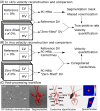
Accelerated dual-venc 4D flow MRI with variable high-venc spatial resolution for neurovascular applications
- PMID: 35754143
- PMCID: PMC9392495
- DOI: 10.1002/mrm.29306
Accelerated dual-venc 4D flow MRI with variable high-venc spatial resolution for neurovascular applications
Abstract
Purpose: Dual-velocity encoded (dual-venc or DV) 4D flow MRI achieves wide velocity dynamic range and velocity-to-noise ratio (VNR), enabling accurate neurovascular flow characterization. To reduce scan time, we present interleaved dual-venc 4D Flow with independently prescribed, prospectively undersampled spatial resolution of the high-venc (HV) acquisition: Variable Spatial Resolution Dual Venc (VSRDV).
Methods: A prototype VSRDV sequence was developed based on a Cartesian acquisition with eight-point phase encoding, combining PEAK-GRAPPA acceleration with zero-filling in phase and partition directions for HV. The VSRDV approach was optimized by varying z, the zero-filling fraction of HV relative to low-venc, between 0%-80% in vitro (realistic neurovascular model with pulsatile flow) and in vivo (n = 10 volunteers). Antialiasing precision, mean and peak velocity quantification accuracy, and test-retest reproducibility were assessed relative to reference images with equal-resolution HV and low venc (z = 0%).
Results: In vitro results for all z demonstrated an antialiasing true positive rate at least 95% for = 2 and 5, with no linear relationship to z (p = 0.62 and 0.13, respectively). Bland-Altman analysis for z = 20%, 40%, 60%, or 80% versus z = 0% in vitro and in vivo demonstrated no bias >1% of venc in mean or peak velocity values at any . In vitro mean and peak velocity, and in vivo peak velocity, had limits of agreement within 15%.
Conclusion: VSRDV allows up to 34.8% scan time reduction compared to PEAK-GRAPPA accelerated DV 4D Flow MRI, enabling large spatial coverage and dynamic range while maintaining VNR and velocity measurement accuracy.
Keywords: 4D flow; acceleration; acquisition; dual-venc; neurovascular.
© 2022 The Authors. Magnetic Resonance in Medicine published by Wiley Periodicals LLC on behalf of International Society for Magnetic Resonance in Medicine.
Conflict of interest statement
Dr. Jianing Pang is an employee of Siemens Medical Solutions USA. Dr. Michael Markl receives research support from Siemens and Circle Cardiovascular Imaging.
Figures


Similar articles
-
Accelerated dual-venc 4D flow MRI for neurovascular applications.J Magn Reson Imaging. 2017 Jul;46(1):102-114. doi: 10.1002/jmri.25595. Epub 2017 Feb 2. J Magn Reson Imaging. 2017. PMID: 28152256 Free PMC article.
-
Dual-Venc acquisition for 4D flow MRI in aortic stenosis with spiral readouts.J Magn Reson Imaging. 2020 Jul;52(1):117-128. doi: 10.1002/jmri.27004. Epub 2019 Dec 18. J Magn Reson Imaging. 2020. PMID: 31850597 Free PMC article.
-
Efficient triple-VENC phase-contrast MRI for improved velocity dynamic range.Magn Reson Med. 2020 Feb;83(2):505-520. doi: 10.1002/mrm.27943. Epub 2019 Aug 18. Magn Reson Med. 2020. PMID: 31423646 Free PMC article.
-
Quantification of peak blood flow velocity at the cardiac valve and great thoracic vessels by four-dimensional flow and two-dimensional phase-contrast MRI compared with echocardiography: a systematic review and meta-analysis.Clin Radiol. 2021 Nov;76(11):863.e1-863.e10. doi: 10.1016/j.crad.2021.07.011. Epub 2021 Aug 14. Clin Radiol. 2021. PMID: 34404516
-
From 2D to 4D Phase-Contrast MRI in the Neurovascular System: Will It Be a Quantum Jump or a Fancy Decoration?J Magn Reson Imaging. 2022 Feb;55(2):347-372. doi: 10.1002/jmri.27430. Epub 2020 Nov 24. J Magn Reson Imaging. 2022. PMID: 33236488 Review.
Cited by
-
Hemodynamics of distal cerebral arteries are associated with functional outcomes in symptomatic ischemic stroke in middle cerebral artery territory: A four-dimensional flow cardiovascular magnetic resonance study.J Cardiovasc Magn Reson. 2025 Summer;27(1):101857. doi: 10.1016/j.jocmr.2025.101857. Epub 2025 Feb 10. J Cardiovasc Magn Reson. 2025. PMID: 39938618 Free PMC article.
-
Motion-corrected 4D-Flow MRI for neurovascular applications.Neuroimage. 2022 Dec 1;264:119711. doi: 10.1016/j.neuroimage.2022.119711. Epub 2022 Oct 25. Neuroimage. 2022. PMID: 36307060 Free PMC article.
-
Four-dimensional flow MRI for quantitative assessment of cerebrospinal fluid dynamics: Status and opportunities.NMR Biomed. 2024 Jul;37(7):e5082. doi: 10.1002/nbm.5082. Epub 2023 Dec 20. NMR Biomed. 2024. PMID: 38124351 Free PMC article. Review.
-
Simultaneous Assessment of Arterial Pulsatility Across Multiple Segments of Cerebral Arteries Using Multiband Dual-VENC Phase-Contrast MRI.NMR Biomed. 2025 Sep;38(9):e70099. doi: 10.1002/nbm.70099. NMR Biomed. 2025. PMID: 40660880 Free PMC article.
References
-
- Schnell S, Ansari SA, Vakil P, et al. Characterization of cerebral aneurysms using 4D FLOW MRI. J Cardiovasc Magn Reson. 2012;14:W2.
-
- Orita E, Murai Y, Sekine T, et al. Four‐dimensional flow MRI analysis of cerebral blood flow before and after high‐flow extracranial–intracranial bypass surgery with internal carotid artery ligation. Neurosurgery. 2018;85:58‐64. - PubMed
-
- Hope TA, Purcell DD, von Morze C, Vigneron DB, Alley MT, Dillon WP. Evaluation of intracranial stenoses and aneurysms with accelerated 4D flow. Magn Reson Imaging. 2010;28(1):41‐46. - PubMed
Publication types
MeSH terms
Grants and funding
LinkOut - more resources
Full Text Sources
Medical

