. 2022 Jun 27;62(12):3107-3122.
doi: 10.1021/acs.jcim.2c00350. Epub 2022 Jun 6.

Singular Interface Dynamics of the SARS-CoV-2 Delta Variant Explained with Contact Perturbation Analysis

Affiliations

Singular Interface Dynamics of the SARS-CoV-2 Delta Variant Explained with Contact Perturbation Analysis

Aria Gheeraert et al. J Chem Inf Model. .

Abstract

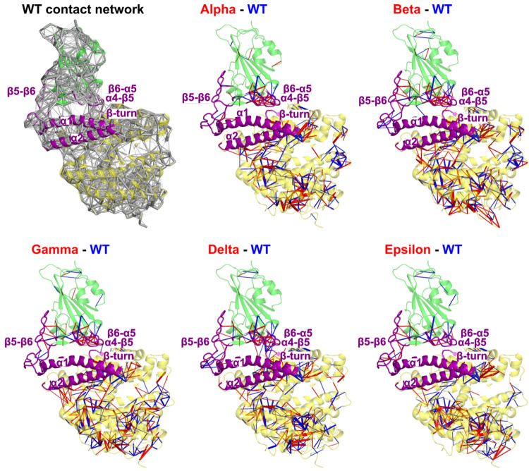
Emerging SARS-CoV-2 variants raise concerns about our ability to withstand the Covid-19 pandemic, and therefore, understanding mechanistic differences of those variants is crucial. In this study, we investigate disparities between the SARS-CoV-2 wild type and five variants that emerged in late 2020, focusing on the structure and dynamics of the spike protein interface with the human angiotensin-converting enzyme 2 (ACE2) receptor, by using crystallographic structures and extended analysis of microsecond molecular dynamics simulations. Dihedral angle principal component analysis (PCA) showed the strong similarities in the spike receptor binding domain (RBD) dynamics of the Alpha, Beta, Gamma, and Delta variants, in contrast with those of WT and Epsilon. Dynamical perturbation networks and contact PCA identified the peculiar interface dynamics of the Delta variant, which cannot be directly imputable to its specific L452R and T478K mutations since those residues are not in direct contact with the human ACE2 receptor. Our outcome shows that in the Delta variant the L452R and T478K mutations act synergistically on neighboring residues to provoke drastic changes in the spike/ACE2 interface; thus a singular mechanism of action eventually explains why it dominated over preceding variants.


Conflict of interest statement

The authors declare no competing financial interest.

Figures

Figure 1
Figure 1
(A) Static amino acid network of the WT. (B–D) Static perturbation network, using the WT (PDB 6M0J) as reference network, at threshold 5 for (B) Alpha (PDB 7EKF, (C) Beta (PDB 7EKG), and (D) Gamma (PDB 7EKC).
Figure 2
Figure 2
Projection of the frames corresponding to the final 400 ns of simulation for the six studied complexes in the two dPCA eigenvector dimensions with (A) contour plots representing a kernel density estimate of the population of each complex and (B) a scatter plot representing the three main clusters obtained through Ward’s minimum variance method. Representation of the influence (as cylinders with a width proportional to the influence) of each dihedral angle in the PC1 (C) and PC2 (D) eigenvectors on the spike RBD (green)/ACE2 (yellow) complex. The α4−β5, β5−β6, and β6−α5 loops are highlighted in purple.
Figure 3
Figure 3
Complete perturbation network between each variant and the WT. The spike RBD (green)/ACE2 (yellow) complex is represented in cartoon representation. Stronger contacts are represented in the WT by a blue edge and in the variants by a red edge. Edge width is proportional to the weight.
Figure 4
Figure 4
(A) Perturbation networks using a threshold value of 5 between the WT RDB (green)/ACE2 (yellow) complex and its mutants (Alpha, Beta, Gamma, Delta, and Epsilon). Stronger contacts are represented in the WT by a blue edge and in the variants by a red edge. Edge width is proportional to the weight and visualization factor, the same for each variant. (B) Average number of interresidual atomic contacts in all pairs at the interface (labeled in the WT residue name) with more than five contacts in at least one simulation.
Figure 5
Figure 5
Projection of the frames corresponding to the final 400 ns of simulation for the six studied complexes in the two cPCA eigenvector dimensions, with (A) terrain lines representing a kernel density estimate of the population of each complex. Network representation of the influence (as cylinders with a width proportional to the influence) of each contact in the PC1 (B) and PC2 (C) and PC2–PC1 (D) eigenvectors projected on the spike RBD (green)/ACE2 (yellow) WT complex. Blue edges show a negative contribution to the principal component, while red edges show a positive contribution to the principal component. Contacts with a contribution of less than 1% to the eigenvector were discarded.
Figure 6
Figure 6
(a–f) Representative MD snapshots of some contacts in the different models emphasized by contact analysis. (g) Summary of the cross-talk between mutated residue T478 and the β6−α5 loop.

References

    1. Wang Q.; Zhang Y.; Wu L.; Niu S.; Song C.; Zhang Z.; Lu G.; Qiao C.; Hu Y.; Yuen K.-Y.; Wang Q.; Zhou H.; Yan J.; Qi J. Structural and functional basis of SARS-CoV-2 entry by using human ACE2. Cell 2020, 181, 894–904. 10.1016/j.cell.2020.03.045. - DOI - PMC - PubMed
    1. Walls A. C.; Park Y.-J.; Tortorici M. A.; Wall A.; McGuire A. T.; Veesler D. Structure, function, and antigenicity of the SARS-CoV-2 spike glycoprotein. Cell 2020, 181, 281–292. 10.1016/j.cell.2020.02.058. - DOI - PMC - PubMed
    1. Lan J.; Ge J.; Yu J.; Shan S.; Zhou H.; Fan S.; Zhang Q.; Shi X.; Wang Q.; Zhang L.; Wang X. Structure of the SARS-CoV-2 spike receptor-binding domain bound to the ACE2 receptor. Nature 2020, 581, 215–220. 10.1038/s41586-020-2180-5. - DOI - PubMed
    1. Yan R.; Zhang Y.; Li Y.; Xia L.; Guo Y.; Zhou Q. Structural basis for the recognition of SARS-CoV-2 by full-length human ACE2. Science 2020, 367, 1444–1448. 10.1126/science.abb2762. - DOI - PMC - PubMed
    1. Castillo A. E.; Parra B.; Tapia P.; Acevedo A.; Lagos J.; Andrade W.; Arata L.; Leal G.; Barra G.; Tambley C.; Tognarelli J.; Bustos P.; Ulloa S.; Fasce R.; Fernández J. Phylogenetic analysis of the first four SARS-CoV-2 cases in Chile. J. Med. Virol. 2020, 92, 1562–1566. 10.1002/jmv.25797. - DOI - PMC - PubMed

Substances

Supplementary concepts