Case Reports
. 2022 May 2;35(4):560-561.
doi: 10.1080/08998280.2022.2068945. eCollection 2022.

A solitary angiokeratoma on the toe of a teenaged girl

Affiliations
Case Reports

A solitary angiokeratoma on the toe of a teenaged girl

Jess Hatfield et al. Proc (Bayl Univ Med Cent). .

Abstract

An angiokeratoma is a benign vascular lesion that appears as one or more red to black papules with a verrucous surface. Histologically, it is defined by ectatic, thin-walled vessels in the papillary dermis, acanthosis with elongated rete ridges, and compact hyperkeratosis. Solitary angiokeratoma is one of five defined subtypes of angiokeratoma. We report the case of an 18-year-old woman with a "wart" that had been present for many years. After treatment with several over-the-counter wart therapies, several rounds of paring plus cryotherapy, and Candida antigen injections failed, a shave biopsy was taken to remove the lesion, and histopathologic examination was consistent with an angiokeratoma. This case demonstrates the importance of considering angiokeratoma in the differential diagnosis of a wart, particularly one recalcitrant to treatment.

Keywords: Angiokeratoma; dermatology; dermatopathology; wart.


Figures

Figure 1.
Figure 1.
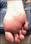
Solitary angiokeratoma on the plantar aspect of the second toe.
Figure 2.
Figure 2.
Solitary angiokeratoma on the plantar aspect of the second toe, shave biopsy. (a) Hematoxylin and eosin staining, 2×. (b–d) Immunohistochemical stains, 5×: (b) D2-40 is partially positive within the endothelial lining. (c) GLUT-1 and (d) WT-1 are negative.

References

    1. Schiller PI, Itin PH.. Angiokeratomas: an update. Dermatology. 1996;193(4):275–282. doi:10.1159/000246270. - DOI - PubMed
    1. Imperial R, Helwig EB.. Angiokeratoma. A clinicopathological study. Arch Dermatol. 1967;95(2):166–175. doi:10.1001/archderm.95.2.166. - DOI - PubMed
    1. Trindade F, Torrelo A, Kutzner H, Requena L, Tellechea Ó, Colmenero I.. An immunohistochemical study of angiokeratomas of children. Am J Dermatopathol. 2014;36(10):796–799. doi:10.1097/dad.0000000000000224. - DOI - PubMed
    1. Wang L, Yuan W, Geng S, et al. . Expression of lymphatic markers in angiokeratomas. J Cutan Pathol. 2014;41(7):576–581. doi:10.1111/cup.12349. - DOI - PubMed
    1. Prindaville B, Lawrence H, Singh V, Nopper AJ, Horii KA.. Immunohistochemical staining of pediatric solitary angiokeratomas. Pediatr Dermatol. 2017;34(2):211–213. doi:10.1111/pde.13089. - DOI - PubMed

Publication types

LinkOut - more resources