Case Reports
. 2022 Jun 10:9:916817.
doi: 10.3389/fmed.2022.916817. eCollection 2022.

Pulmonary Actinomyces graevenitzii Infection: Case Report and Review of the Literature

Affiliations
Case Reports

Pulmonary Actinomyces graevenitzii Infection: Case Report and Review of the Literature

Yuan Yuan et al. Front Med (Lausanne). .

Abstract

Background: Pulmonary actinomycosis (PA), a chronic indolent infection, is a diagnostic challenge. Actinomyces graevenitzii is a relatively rare Actinomyces species isolated from various clinical samples.

Case presentation: A 47-year-old patient presented with a 3-month history of mucopurulent expectoration and dyspnea and a 3-day history of fever up to 39.0°C. He had dental caries and a history of alcoholism. Computed tomography (CT) images of the chest revealed a consolidation shadow in the right upper and middle lobes, with necrosis containing foci of air. Actinomyces graevenitzii was isolated from bronchoalveolar lavage fluid (BALF) culture and was identified by matrix-assisted laser desorption/ionization time-of-flight mass spectrometry. He received treatment with intravenous piperacillin-sulbactam for 10 days and oral amoxicillin-clavulanate for 7 months. His clinical condition had considerably improved. The consolidation shadow was gradually absorbed.

Conclusion: Early diagnosis and treatment of pulmonary actinomycosis are crucial. Bronchoscopy plays a key role in the diagnostic process, and matrix-assisted laser desorption/ionization time-of-flight mass spectrometry (MALDI-TOF/MS) is an accurate tool for Actinomyces identification.

Keywords: Actinomyces graevenitzii; bronchoscopy; consolidation; matrix-assisted laser desorption/ionization time-of-flight mass spectrometry; pulmonary actinomycosis (PA).


Conflict of interest statement

The authors declare that the research was conducted in the absence of any commercial or financial relationships that could be construed as a potential conflict of interest.

Figures

FIGURE 1
FIGURE 1
Chest computed tomography images at admission. A consolidation shadow in the right upper and middle lobes, with necrosis containing foci of air. (A,C) lung window; (B,D) mediastinal window.
FIGURE 2
FIGURE 2
A series of bronchoscopy images. The first bronchoscopy images (A–C), the secondary bronchoscopy images (D–F). (A) The medial and lateral segments of the RML were blocked by purulent yellow secretions. (B) The medial subsegment of the RML was completely obstructed by an endobronchial white necrotized mass. (C) The media subsegment of the RML became unobstructed after suction. (D) The medial and lateral segments of the RML were blocked by purulent yellow secretions. (E) The medial subsegment of the RML was completely obstructed by a an endobronchial white necrotized mass. (F) The media subsegment of the RML became unobstructed after suction. RML: Right middle lobe.
FIGURE 3
FIGURE 3
Gram stain of the bronchoalveolar lavage fluid.
FIGURE 4
FIGURE 4
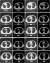
Serial changes on chest computed tomography findings. Chest CT at discharge (A–D). Chest CT at one month’s follow-up (E–H). Chest CT at three months’ follow-up (I–L). Chest CT at five months’ follow-up (M–P). Chest CT at seven months’ follow-up (Q–T). CT: computed tomography.
FIGURE 5
FIGURE 5
Timeline with the most relevant data of the clinical case.

References

    1. Valour F, Sénéchal A, Dupieux C, Karsenty J, Lustig S, Breton P, et al. Actinomycosis: etiology, clinical features, diagnosis, treatment, and management. Infect Drug Resist. (2014) 7:183–97. 10.2147/IDR.S39601 - DOI - PMC - PubMed
    1. Tabarsi P, Yousefi S, Jabbehdari S, Marjani M, Baghaei P. Pulmonary actinomycosis in a patient with AIDS/HCV. J Clin Diagn Res. (2017) 11:OD15–7. 10.7860/JCDR/2017/27593.10092 - DOI - PMC - PubMed
    1. Kheir F, Hamdi T, Khayr W, Latta S. Nonresolving pneumonia. Am J Ther. (2011) 18:e177–9. - PubMed
    1. Crisafulli E, Bernardinello N, Alfieri V, Pellegrino F, Lazzari C, Gnetti L, et al. A pulmonary infection by Actinomyces odontolyticus and Veillonella atypica in an immunocompetent patient with dental caries. Respirol Case Rep. (2019) 7:e00493. 10.1002/rcr2.493 - DOI - PMC - PubMed
    1. Mosimann J, Hany A, Kayser FH. Pulmonary Actinomyces-viscosus infection. Schweiz Med Wochenschr. (1979) 109:720–2. - PubMed

Publication types

LinkOut - more resources