

Optical Coherence Tomography Biomarkers in Predicting Treatment Outcomes of Diabetic Macular Edema After Dexamethasone Implants
- PMID: 35755055
- PMCID: PMC9218219
- DOI: 10.3389/fmed.2022.852022
Optical Coherence Tomography Biomarkers in Predicting Treatment Outcomes of Diabetic Macular Edema After Dexamethasone Implants
Abstract
Purpose: To identify optical coherence tomography (OCT) biomarkers that may predict functional and anatomical outcomes in diabetic macular edema (DME) patients treated with intravitreal dexamethasone (DEX) implant.
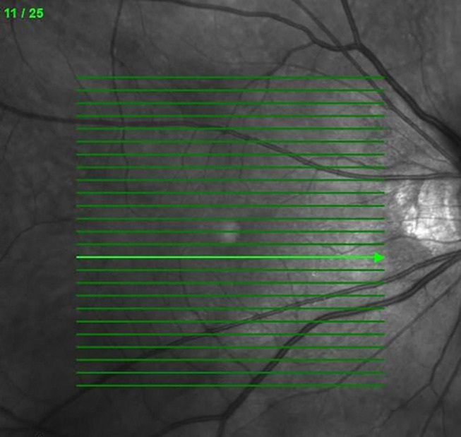
Materials and methods: Sixty-four eyes from 50 patients with DME were enrolled. Best-corrected visual acuity (BCVA) and OCT biomarkers including central retinal thickness (CRT), subretinal fluid (SRF), intraretinal cysts (IRC), ellipsoid zone disruption (EZD), disorganization of retinal inner layers (DRIL), hard exudate (HE), hyperreflective foci (HRF), epiretinal membrane (ERM), and vitreomacular interface (VMI) changes were evaluated at baseline and at 3, 6, and 12 months after treatment. Multiple logistic analysis was performed to evaluate each OCT biomarker as a predictive factor for functional and anatomical improvement at the end of treatment.
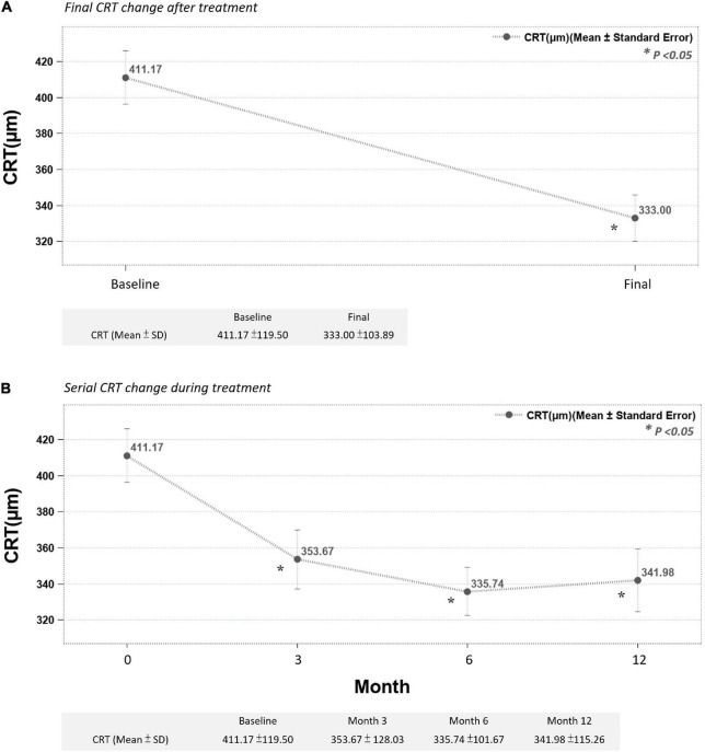
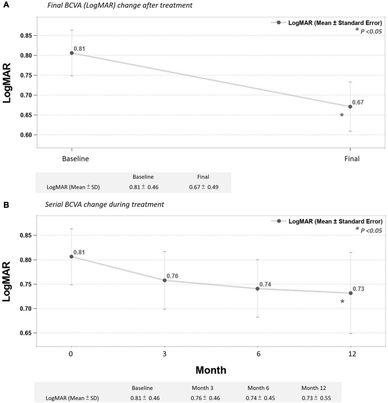
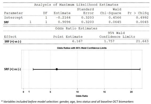
Results: The presence of SRF at baseline was associated with a favorable outcome, with CRT improving by more than 100 μm after treatment from multivariate logistic regression analysis [odds ratio 6.16 (1.75-21.6)]. In addition, baseline SRF predicted a greater CRT improvement from multiple regression analysis (model R-square 0.11, p = 0.006). The reduction of DRIL, SRF, LONLC, IRC, and EZD were correlated with better CRT improvement (more than 100 μm) (P < 0.05). SRF and EZD recovery can also predict better visual prognosis (P < 0.05).
Conclusion: OCT biomarkers can be used to predict who may benefit the most after DEX treatment. We suggest that the DEX implant should be considered as a first line treatment in DME patients with SRF.
Keywords: diabetic macular edema (DME); disorganization of retinal inner layers; hyperreflective foci; intravitreal dexamethasone implant; optical coherence tomography biomarkers; subretinal fluid.
Copyright © 2022 Huang, Chang, Meng, Lin, Lai, Hsia, Chen, Tien, Bair, Lin, Chen and Tsai.
Conflict of interest statement
H-SC was employed by NephroCare Ltd. The remaining authors declare that the research was conducted in the absence of any commercial or financial relationships that could be construed as a potential conflict of interest.
Figures
References
-
- International Diabetes Federation. IDF Diabetes Atlas. 8th ed. Brussels: International Diabetes Federation; (2017).
LinkOut - more resources
Full Text Sources
Research Materials
Miscellaneous