Case Reports
. 2022 Jun 17;17(9):2968-2971.
doi: 10.1016/j.radcr.2022.05.053. eCollection 2022 Sep.

Co-existing fast CSF leaks and CSF-venous fistulas on dynamic CT myelography

Affiliations
Case Reports

Co-existing fast CSF leaks and CSF-venous fistulas on dynamic CT myelography

Ajay A Madhavan et al. Radiol Case Rep. .

Abstract

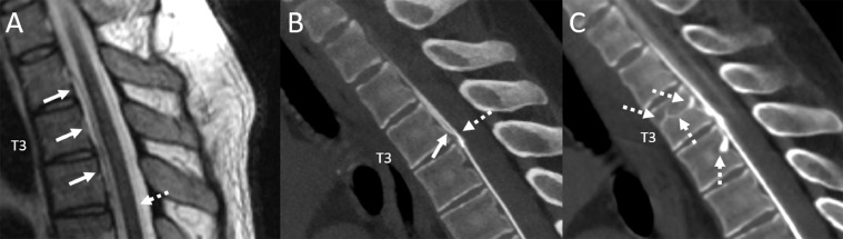
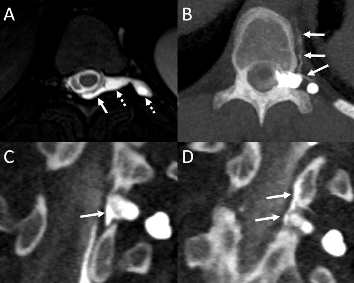
Spontaneous intracranial hypotension can be caused by spinal dural tears or CSF-venous fistulas. It is rare for patients to have more than one type of leak at any given time. Here, we illustrate 3 examples of dural tears that co-existed with CSF-venous fistulas, with both being seen on dynamic CT myelography. To our knowledge, coexistent CSF-venous fistulas and dural tears have not been previously illustrated on dynamic CT myelography, even though this is one of the most commonly used modalities to work-up patients with CSF leaks. We discuss the clinical importance of the rare co-occurrence of these leaks with regard to diagnosis and treatment, as well as implications for understanding and classifying CSF leaks.

Keywords: CSF-venous fistula; CVF, CSF-venous fistula; Dynamic CT myelography; Extradural fluid collection; Fast CSF leak; Lateral decubitus; SIH, spontaneous intracranial hypotension; SLEC, spinal longitudinal extradural collection.


Figures

Fig 1
Fig. 1
Sagittal T2W thoracic spine MRI shows a thin ventral epidural fluid collection spanning T2 through T4 (A, arrows). Sagittal reconstructed image from a prone dynamic CT myelogram (B) demonstrates contrast leak into the ventral fluid collection at the T3-T4 interspace, compatible with fast leak from a ventral dural tear (B, dashed arrow), with superior flow of leaked epidural contrast (B, solid arrow). Sagittal image from a subsequent scan obtained one minute later shows a complex CSF-venous fistula involving the basivertebral veins at T3 and T4 (C, dashed arrows).
Fig 2
Fig. 2
Sagittal T2W MRI demonstrates a thin ventral extradural fluid collection (A, arrows). Sagittal reconstructed image from a subsequent prone dynamic CT myelogram shows a ventral leak at the T8-T9 interspace with clearly distinct epidural contrast (B, solid arrow) and intrathecal contrast (B, dashed arrow). Axial image from the same exam demonstrates a T6 CSF-venous fistula with opacification of intraosseous veins (C, dashed arrows), as well as the right paraspinal vein (C, solid arrow).
Fig 3
Fig. 3
Axial T2W MR image shows a large left T10 meningeal diverticulum (A, dashed arrows) and adjacent dorsal epidural fluid collection (A, solid arrow). Multiple consecutive axial (A) and coronal reformatted (C-D) images from a left lateral decubitus dynamic CT myelogram show a left T10 CSF-venous fistula arising from the meningeal diverticulum (B, arrows). There is an additional adjacent dural leak from the proximal nerve root sleeve along the same left T10 diverticulum (C, solid arrows). Epidural contrast extends superiorly on a subsequently obtained image (D, solid arrows).

References

    1. Farb R.I., Nicholson P.J., Peng P.W., Massicotte E.M., Lay C., Krings T., et al. Spontaneous Intracranial Hypotension: A Systematic Imaging Approach for CSF Leak Localization and Management Based on MRI and Digital Subtraction Myelography. AJNR Am J Neuroradiol. 2019;40(4):745–753. - PMC - PubMed
    1. Schievink W.I., Maya M.M., Jean-Pierre S., Nuno M., Prasad R.S., Moser F.G. A classification system of spontaneous spinal CSF leaks. Neurology. 2016;87(7):673–679. - PubMed
    1. Kim D.K., Carr C.M., Benson J.C., Diehn F.E., Lehman V.T., Liebo G.B., et al. Diagnostic Yield of Lateral Decubitus Digital Subtraction Myelogram Stratified by Brain MRI Findings. Neurology. 2021;96(9) e1312-e8. - PubMed
    1. Mamlouk M.D., Ochi R.P., Jun P., Shen P.Y. Decubitus CT Myelography for CSF-Venous Fistulas: A Procedural Approach. AJNR Am J Neuroradiol. 2021;42(1):32–36. - PMC - PubMed
    1. Schievink W.I., Maya M.M., Moser F., Prasad R., Wadhwa V., Cruz R., et al. Multiple Spinal CSF Leaks in Spontaneous Intracranial Hypotension: Do They Exist? Neurol Clin Pract. 2021;11(5) e691-e7. - PMC - PubMed

Publication types

LinkOut - more resources