Review
. 2022 Mar 18;3(6):648-697.
doi: 10.1039/d2cb00017b. eCollection 2022 Jun 8.

Perspectives on conformationally constrained peptide nucleic acid (PNA): insights into the structural design, properties and applications

Affiliations
Review

Perspectives on conformationally constrained peptide nucleic acid (PNA): insights into the structural design, properties and applications

Chaturong Suparpprom et al. RSC Chem Biol. .

Abstract

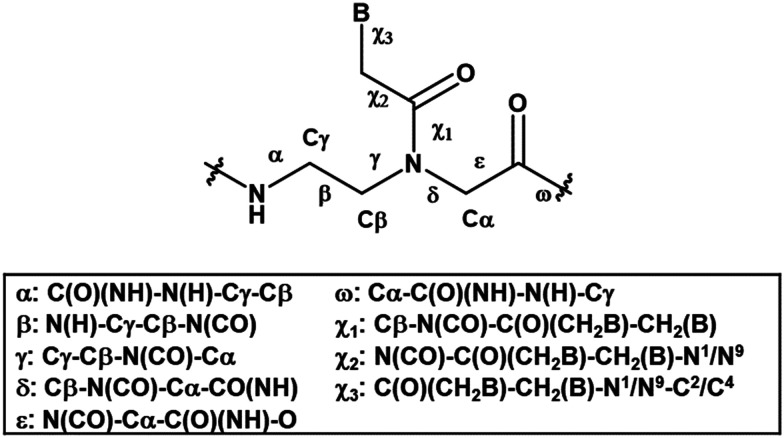
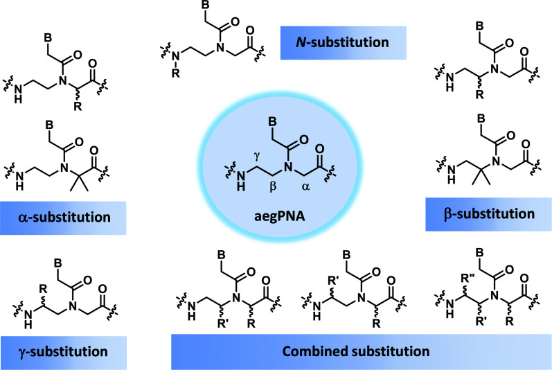
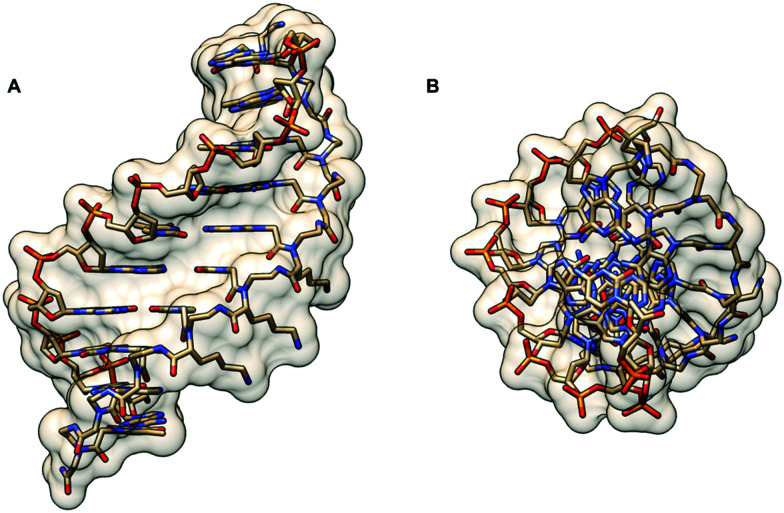
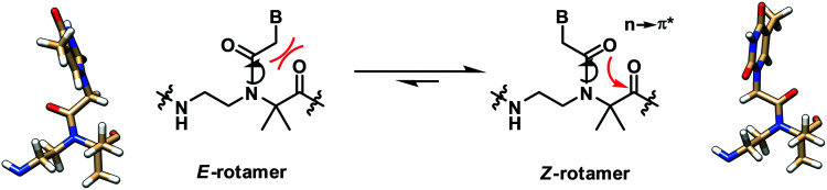
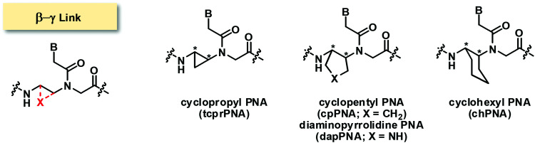
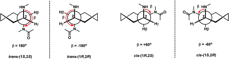
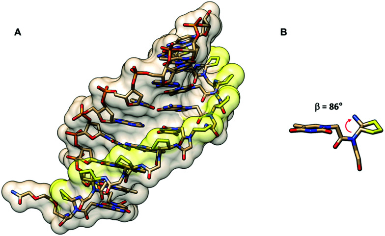
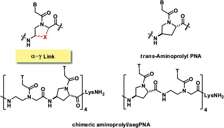
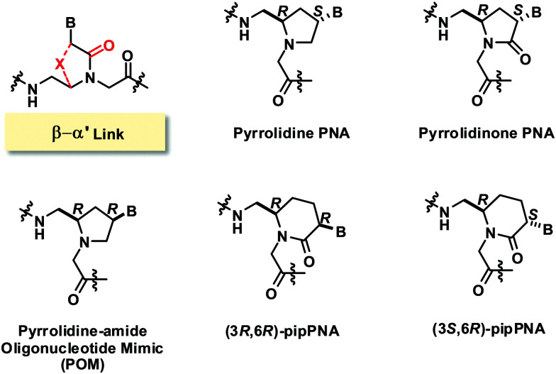
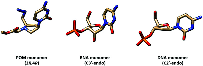
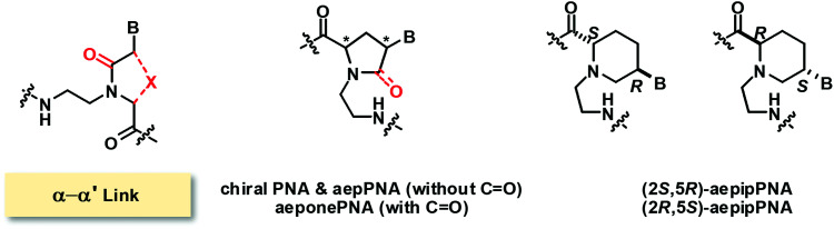
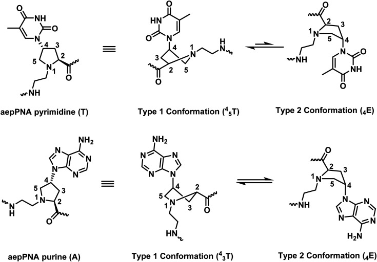
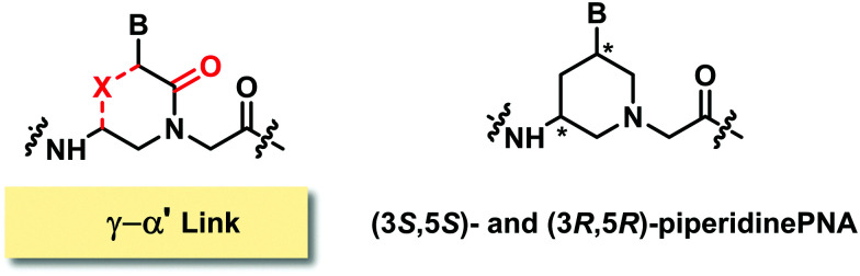
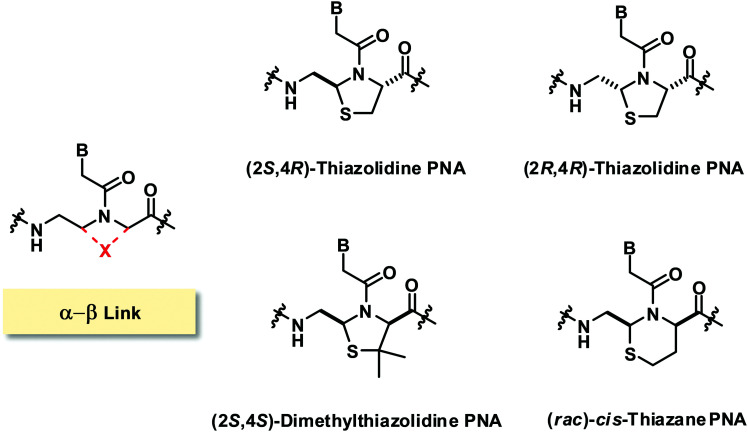
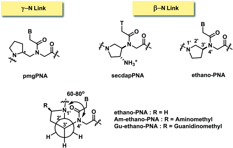
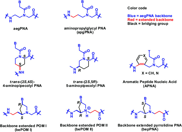
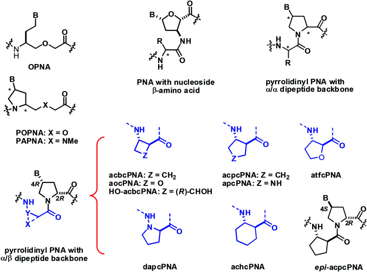
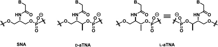
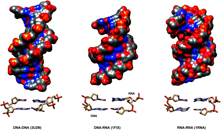
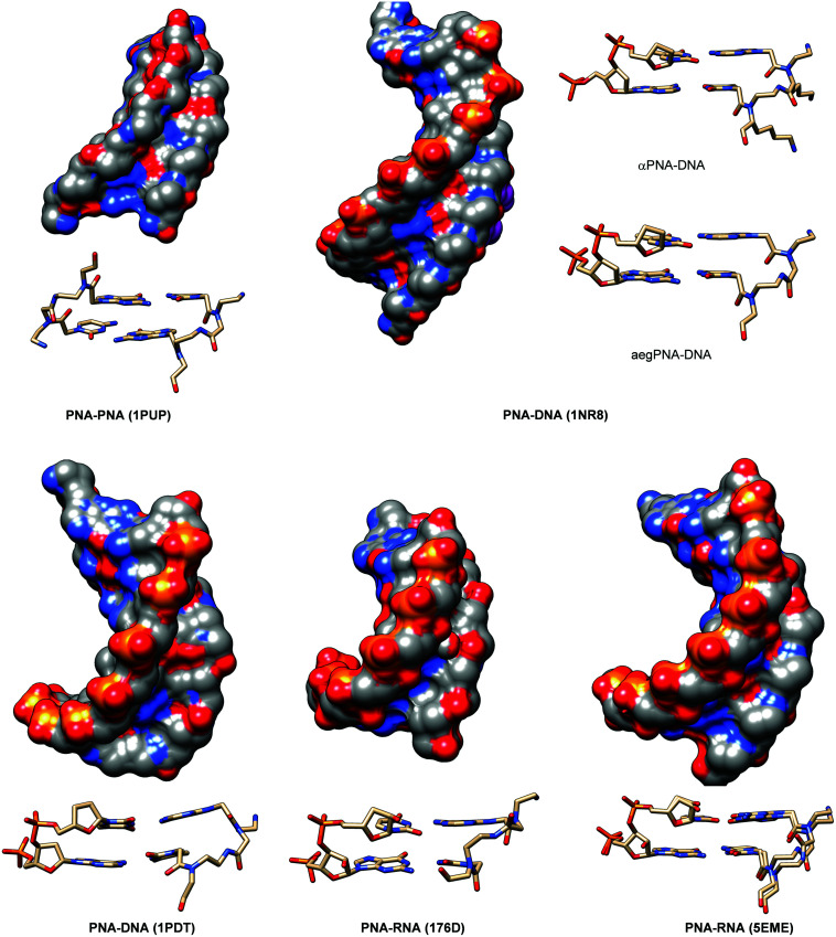
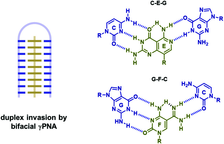
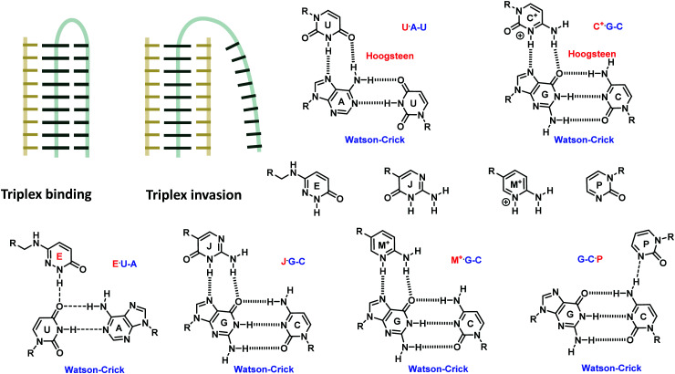
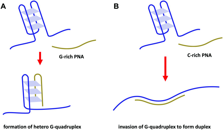
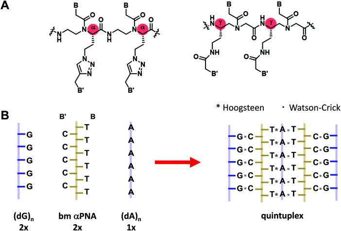
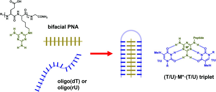
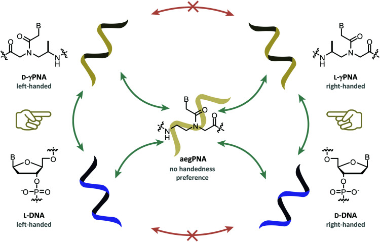
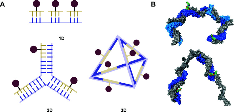
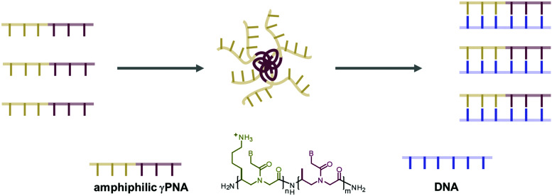
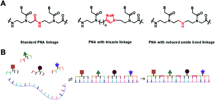
Peptide nucleic acid or PNA is a synthetic DNA mimic that contains a sequence of nucleobases attached to a peptide-like backbone derived from N-2-aminoethylglycine. The semi-rigid PNA backbone acts as a scaffold that arranges the nucleobases in a proper orientation and spacing so that they can pair with their complementary bases on another DNA, RNA, or even PNA strand perfectly well through the standard Watson-Crick base-pairing. The electrostatically neutral backbone of PNA contributes to its many unique properties that make PNA an outstanding member of the xeno-nucleic acid family. Not only PNA can recognize its complementary nucleic acid strand with high affinity, but it does so with excellent specificity that surpasses the specificity of natural nucleic acids and their analogs. Nevertheless, there is still room for further improvements of the original PNA in terms of stability and specificity of base-pairing, direction of binding, and selectivity for different types of nucleic acids, among others. This review focuses on attempts towards the rational design of new generation PNAs with superior performance by introducing conformational constraints such as a ring or a chiral substituent in the PNA backbone. A large collection of conformationally rigid PNAs developed during the past three decades are analyzed and compared in terms of molecular design and properties in relation to structural data if available. Applications of selected modified PNA in various areas such as targeting of structured nucleic acid targets, supramolecular scaffold, biosensing and bioimaging, and gene regulation will be highlighted to demonstrate how the conformation constraint can improve the performance of the PNA. Challenges and future of the research in the area of constrained PNA will also be discussed.


Conflict of interest statement

There are no conflicts of interest to declare.

Figures

Fig. 1
Fig. 1. The structure of aegPNA with definition of backbone atom positions and torsional angles.
Fig. 2
Fig. 2. Possible sites for modification of aegPNA without forming a cyclic structure as part of the backbone.
Fig. 3
Fig. 3. X-Ray structure of the αPNA·DNA duplex (1NR8): (A) side view from the minor groove side and (B) top view from the 3′/N-side along the helix axis.
Fig. 4
Fig. 4. Interconversion of the E- and Z-rotamers in the α-gem-dimethylated PNA monomer.
Fig. 5
Fig. 5. A model of the βPNA structure showing a different degree of steric clash between the β-(S)- or β-(R)-methyl groups and the nucleobase side arm (reprinted from ref. , Copyright (2011), with permission from Elsevier).
Fig. 6
Fig. 6. The gauche conformation of the N(H)–Cγ–Cβ–N(CO) groups necessitates pre-organization into the right-handed helix in β-(S)-methyl PNA. The structure is viewed along the Cγ–Cβ axis.
Fig. 7
Fig. 7. Interconversion of the E- and Z-rotamers in the β-gem-dimethylated PNA monomer.
Fig. 8
Fig. 8. The gauche conformation of the N(H)–Cγγ–Cβ–N(CO) groups necessitates pre-organization into the right-handed helix in γ-(S)-methyl PNA. The structure is viewed along the Cγ–Cβ axis (adapted with permission from ref. . Copyright 2006 American Chemical Society).
Fig. 9
Fig. 9. (A) X-Ray structure of the γPNA·DNA duplex (3PA0) viewed from the minor groove side. (B) Superposition of the PNA strand from the γPNA·DNA duplex (cyan), aegPNA·DNA duplex (green), and single-stranded γPNA dimer (blue) (reprinted with permission from ref. . Copyright 2010 American Chemical Society).
Fig. 10
Fig. 10. Interconversion of the E- and Z-rotamers in the γ-gem-dimethylated PNA monomer.
Fig. 11
Fig. 11. X-Ray structure of the N-Me PNA·PNA duplex (1QPY) viewed from the minor groove side with the different orientation of the backbone carbonyl groups of the N-Me and normal aegPNA monomers shown.
Fig. 12
Fig. 12. (A) Structure of β,γ-modified PNA. (B) The proposed different conformation of the OH and OMe substituted PNA (reproduced from ref. 57 with permission from the Royal Society of Chemistry).
Fig. 13
Fig. 13. Possible sites for modification of aegPNA by forming a cyclic structure as part of the backbone.
Fig. 14
Fig. 14. Structures of modified PNA with (β–γ)-linkage.
Fig. 15
Fig. 15. Structures of cyclohexane-modified PNA with (β–γ)-linkage.
Fig. 16
Fig. 16. (A) X-Ray structure of the trans-(S,S)-cpPNA·DNA duplex (7KZL) viewed from the minor groove side. (B) Torsional angle β of the diaminocyclopentane unit from the X-ray structure of the cpPNA·DNA duplex. The cyclopentyl modification of the PNA backbone is shown in yellow.
Fig. 17
Fig. 17. Structures of modified PNA with (α–γ)-linkage.
Fig. 18
Fig. 18. Structures of modified PNA with (β–α′)-linkage.
Fig. 19
Fig. 19. Comparison of conformation of the pyrrolidine ring in the POM monomer and sugar ring puckering in generic RNA and DNA structures.
Fig. 20
Fig. 20. Structures of modified PNA with (α–α′)-linkage.
Fig. 21
Fig. 21. The base-dependency of the conformation of the pyrrolidine ring in aepPNA (reprinted from ref. , Copyright (2010), with permission from Elsevier).
Fig. 22
Fig. 22. Structures of modified PNA with (γ–α′)-linkage.
Fig. 23
Fig. 23. Structures of modified PNA with (α−β)-linkage.
Fig. 24
Fig. 24. Structures of modified PNA with (γ-N)- and (β-N)-linkages.
Fig. 25
Fig. 25. Structures of constrained aegPNA with extended backbones.
Fig. 26
Fig. 26. Structures of conformationally constrained PNA that does not follow the generic aegPNA template.
Fig. 27
Fig. 27. (A) Structures of pyrrolidinyl PNA homologs. (B) Torsional angle N(H)–Cα–Cβ–C(O) of (1S,2S)-ACPC in ACPC PNA. C) Native torsional angles of cyclic β-amino acid oligomers are obtained from the literature (adapted with permission from ref. . Copyright 2012 American Chemical Society).
Fig. 28
Fig. 28. Structures of SNA, d-aTNA and l-aTNA.
Fig. 29
Fig. 29. Three-dimensional structures of standard DNA·DNA, DNA·RNA and RNA·RNA duplexes, with details of the monomer conformation and base-pairing.
Fig. 30
Fig. 30. Three-dimensional structures of PNA·PNA, PNA·DNA and PNA·RNA duplexes, with details of the monomer conformation and base-pairing.
Fig. 31
Fig. 31. Three-dimensional structures of γPNA·PNA and cpPNA·DNA duplexes, with details of the monomer conformation and base-pairing. The backbone substituents are highlighted in yellow.
Fig. 32
Fig. 32. Comparison of Tm values of selected PNA·DNA duplexes (A and B) and PNA·RNA duplexes (C). Color code for X in the DNA/RNA strand: A (blue), C (red), G (green), T/U (purple). γPNA(1) = LSer γPNA; γPNA(2) = LminiPEG (LMP) γPNA. Tm data were taken from ref. , , , and (adapted with permission from ref. . Copyright 2015 American Chemical Society).
Fig. 33
Fig. 33. Various modes of targeting DNA duplexes (blue) by PNA (gold).
Fig. 34
Fig. 34. (A) Watson–Crick/Hoogsteen base triplets involved in the triplex formation between PNA (gold) and DNA (blue). (B) A pseudocomplementary base-pairing scheme for A·T pairs.
Fig. 35
Fig. 35. Invasion of DNA duplexes by bifacial PNA.
Fig. 36
Fig. 36. Targeting RNA duplexes (gray) by PNA (gold) via triplex formation and invasion, and the structures of the modified base that facilitate the triplex binding mode.
Fig. 37
Fig. 37. Targeting DNA G-quadruplexes (blue) by PNA (gold) by hetero G-quadruplex formation (A) or duplex formation (B).
Fig. 38
Fig. 38. (A) Structures of bimodal αPNA and γPNA. (B) Spontaneous assembly of bimodal PNA (gold) with DNA (blue) to form complex structures.
Fig. 39
Fig. 39. Structures of bifacial PNA containing the melamine base (gold) and its interaction with thymine in DNA (blue) or uracil in RNA strands to form a stable triplex structure (adapted with permission from ref. . Copyright 2012 American Chemical Society).
Fig. 40
Fig. 40. Orthogonality pairing in γPNA (gold) and DNA (blue). Only γPNA and DNA with the same handedness can self-pair and cross-pair. The achiral aegPNA pairs indiscriminately with all; thus it can act as a mediator to transmit the information between these orthogonal nucleic acids.
Fig. 41
Fig. 41. (A) Schematic representation of using PNA for multivalent display of ligands on various DNA scaffolds. (B) A model showing the spatial arrangement of two N-acetyllactosamine (LacNAc) ligands in two different multivalent constructs derived from γPNA (blue) and DNA (gray) (reproduced from ref. with permission from the Royal Society of Chemistry).
Fig. 42
Fig. 42. The self-assembly of amphiphilic γPNA probes is modulated by γPNA·DNA/RNA duplex formation (adapted with permission from ref. . Copyright 2019 American Chemical Society).
Fig. 43
Fig. 43. (A) Some chemistry useful for template-directed PNA oligomerization. (B) The self-assembly and oligomerization of functionalized PNA probes on a DNA template.
Fig. 44
Fig. 44. Dynamic sequence adaptive behavior of thioester peptide nucleic acids (tPNA).
Fig. 45
Fig. 45. Examples of biosensing strategies that took the advantages of the duplex invasion ability of modified PNA probes. (A) Chemiluminescence microbeads γPNA array for the rapid and simultaneous identification of blood pathogens. (B) Nanopore-based sequencing and barcoding of long dsDNA strands by γPNA probes (adapted with permission from ref. . Copyright 2012 American Chemical Society).
Fig. 46
Fig. 46. Examples of strategies for cellular imaging that took the advantages of high affinity modified PNA probes. (A) RNA imaging by templated Staudinger reaction between two PNA probes. (B) Visualization of telomeric DNA by PNA probes. (C) Visualization of carbonic anhydrase IX with the aid of γPNA probes and HCR.
None
Chaturong Suparpprom
None
Tirayut Vilaivan

References

    1. Buccitelli C. Selbach M. mRNAs, proteins and the emerging principles of gene expression control. Nat. Rev. Genet. 2020;21:630–644. doi: 10.1038/s41576-020-0258-4. - DOI - PubMed
    1. Chaudhary N. Weissman D. Whitehead K. A. mRNA Vaccines for infectious diseases: Principles, delivery and clinical translation. Nat. Rev. Drug Discovery. 2021;20:817–838. doi: 10.1038/s41573-021-00283-5. - DOI - PMC - PubMed
    1. Morihiro K. Kasahara Y. Obika S. Biological applications of xeno nucleic acids. Mol. Biosyst. 2017;13:235–245. doi: 10.1039/C6MB00538A. - DOI - PubMed
    1. Duffy K. Arangundy-Franklin S. Holliger P. Modified nucleic acids: Replication, evolution, and next-generation therapeutics. BMC Biolol. 2020;18:112. doi: 10.1186/s12915-020-00803-6. - DOI - PMC - PubMed
    1. Ruiz-Mirazo K. Briones C. de la Escosura A. Prebiotic systems chemistry: New perspectives for the origins of life. Chem. Rev. 2014;114:285–366. doi: 10.1021/cr2004844. - DOI - PubMed