. 2022 Jun 7;7(24):21337-21345.
doi: 10.1021/acsomega.2c03055. eCollection 2022 Jun 21.

Cerium Oxide Nanoparticles with Entrapped Gadolinium for High T 1 Relaxivity and ROS-Scavenging Purposes

Affiliations

Cerium Oxide Nanoparticles with Entrapped Gadolinium for High T 1 Relaxivity and ROS-Scavenging Purposes

Peter Eriksson et al. ACS Omega. .

Abstract

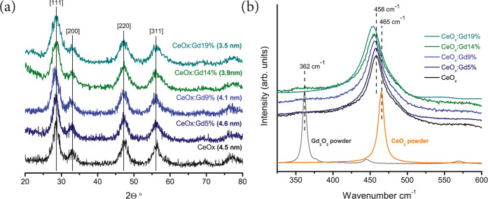
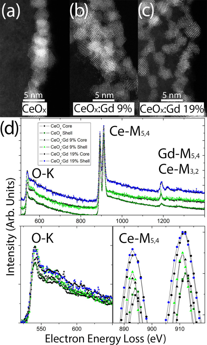
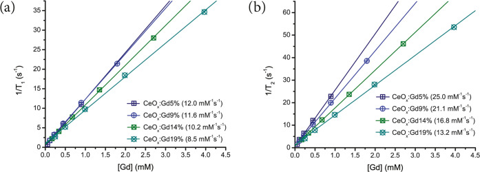
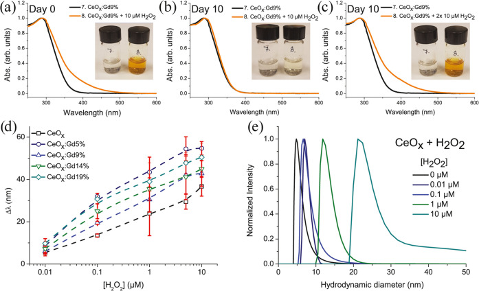
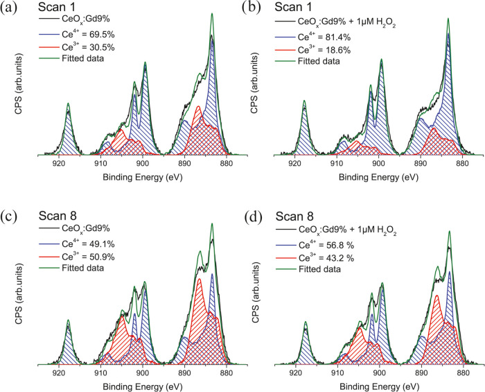
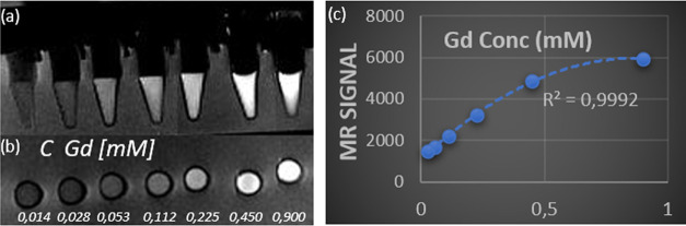
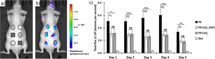
Gadolinium chelates are employed worldwide today as clinical contrast agents for magnetic resonance imaging. Until now, the commonly used linear contrast agents based on the rare-earth element gadolinium have been considered safe and well-tolerated. Recently, concerns regarding this type of contrast agent have been reported, which is why there is an urgent need to develop the next generation of stable contrast agents with enhanced spin-lattice relaxation, as measured by improved T 1 relaxivity at lower doses. Here, we show that by the integration of gadolinium ions in cerium oxide nanoparticles, a stable crystalline 5 nm sized nanoparticulate system with a homogeneous gadolinium ion distribution is obtained. These cerium oxide nanoparticles with entrapped gadolinium deliver strong T 1 relaxivity per gadolinium ion (T 1 relaxivity, r 1 = 12.0 mM-1 s-1) with the potential to act as scavengers of reactive oxygen species (ROS). The presence of Ce3+ sites and oxygen vacancies at the surface plays a critical role in providing the antioxidant properties. The characterization of radial distribution of Ce3+ and Ce4+ oxidation states indicated a higher concentration of Ce3+ at the nanoparticle surfaces. Additionally, we investigated the ROS-scavenging capabilities of pure gadolinium-containing cerium oxide nanoparticles by bioluminescent imaging in vivo, where inhibitory effects on ROS activity are shown.


Conflict of interest statement

The authors declare no competing financial interest.

Figures

Figure 1
Figure 1
(a) X-ray diffraction patterns of CeOx, CeOx:Gd5%, CeOx:Gd9%, CeOx:Gd14%, and CeOx:Gd19%. The calculated grain sizes as determined by the Scherrer equation are given. (b) Raman spectra for CeO2, Gd2O3, CeOx, CeOx:Gd5%, CeOx:Gd9%, CeOx:Gd14%, and CeOx:Gd19%. The Raman spectra are normalized and vertically shifted using a small offset for each spectrum to facilitate line shape comparison. There is an increasing shift of the main Raman peak to lower wavenumbers related to the integration of Gd.
Figure 2
Figure 2
(a–c) High-resolution HAADF-STEM images of the CeOx, CeOx:Gd9%, and CeOx:Gd19% particles, respectively. (d) Vertically shifted core-loss EEL spectra (top) for the same samples, with emphasis on baseline aligned the O-K (bottom left) and vertically shifted Ce-M (bottom right) edges.
Figure 3
Figure 3
Inverse of relaxation times (a) 1/T1 and (b) 1/T2 are shown as a function of the concentration of gadolinium for the prepared Gd-CeNPs. The slope of the fitted linear equations denoted as the r1 (a) and r2 (b) relaxivities are given within the brackets.
Figure 4
Figure 4
Absorbance spectra and inserted images of CeOx:Gd9% untreated and treated with 10 μM H2O2 on (a) day 0, (b) day 10, and (c) on day 10, another 10 μM H2O2 was added to the sample. (d) Plotted red shifts CeOx:Gd0–19% ([Ce] = 10 μg mL–1) treated with increasing concentration of H2O2. (e) Hydrodynamic diameter of CeOx treated with H2O2 (number-weighted distributions). The photos in panels (a)–(c) courtesy of the first author, P. Eriksson. Copyright 2020.
Figure 5
Figure 5
XPS spectra of (a) CeOx:Gd9% scan 1, (b) CeOx:Gd9% treated with 1 μM H2O2 scan 1, (c) CeOx:Gd9% scan 8, and (d) CeOx:Gd9% treated with 1 μM H2O2 scan 1.
Figure 6
Figure 6
T1-weighted MR images of CeOx:Gd9% nanoparticles as a function of Gd concentration: (a) schematic image side view of measured tubes, (b) top view, and (c) measured MR signal (T1-weighted) from the cylindrical volume of 3 mm height and 5 mm diameter.
Figure 7
Figure 7
Antioxidant behavior of CeOx and CeOx:Gd5% nanoparticles in immunocompetent mice. (a) Schematic of subcutaneous injection on the dorsal side of a mouse showing positions of different particle formulations including a mixture of CeOx with PS (hexagonal), a mixture of CeOx:Gd5% with PS (circle), and PS microparticles only (square) as a control. (b) Bioluminescent image of the ROS signal from a representative mouse on day 3, following subcutaneous injection of particles. (c) Quantification of bioluminescent signals from injection sites over a 5 day period. Error bars are standard of the mean (n = 12 injections on 6 mice with 2 replicates per mouse). *, **, ***, **** denote p ≤ 0.05, 0.01, 0.001, and 0.0001, respectively. “ns” denotes nonsignificance, which has p > 0.05.

References

    1. Todd D. J.; Kay J. Nephrogenic systemic fibrosis: An epidemic of gadolinium toxicity. Curr. Rheumatol. Rep. 2008, 10, 195–204. 10.1007/s11926-008-0033-6. - DOI - PubMed
    1. Sharma P.; Brown S.; Walter G.; Santra S.; Moudgil B. Nanoparticles for bioimaging. Adv. Colloid Interface Sci. 2006, 123–126, 471–485. 10.1016/j.cis.2006.05.026. - DOI - PubMed
    1. Ahrén M.; Selegård L.; Klasson A.; Söderlind F.; Abrikossova N.; Skoglund C.; Bengtsson T.; Engström M.; Käll P.-O.; Uvdal K. Synthesis and Characterization of PEGylated Gd2O3 Nanoparticles for MRI Contrast Enhancement. Langmuir 2010, 26, 5753–5762. 10.1021/la903566y. - DOI - PubMed
    1. Ahrén M.; Selegård L.; Söderlind F.; Linares M.; Kauczor J.; Norman P.; Käll P.-O.; Uvdal K. A simple polyol-free synthesis route to Gd2O3 nanoparticles for MRI applications: an experimental and theoretical study. J. Nanopart. Res. 2012, 14, 100610.1007/s11051-012-1006-2. - DOI
    1. Park J. Y.; Baek M. J.; Choi E. S.; Woo S.; Kim J. H.; Kim T. J.; Jung J. C.; Chae K. S.; Chang Y.; Lee G. H. Paramagnetic Ultrasmall Gadolinium Oxide Nanoparticles as Advanced T1 MRI Contrast Agent: Account for Large Longitudinal Relaxivity, Optimal Particle Diameter, and In Vivo T1 MR Images. ACS Nano 2009, 3, 3663–3669. 10.1021/nn900761s. - DOI - PubMed