

Thiol-ene conjugation of VEGF peptide to electrospun scaffolds as potential application for angiogenesis
- PMID: 35755423
- PMCID: PMC9192696
- DOI: 10.1016/j.bioactmat.2022.05.029
Thiol-ene conjugation of VEGF peptide to electrospun scaffolds as potential application for angiogenesis
Abstract
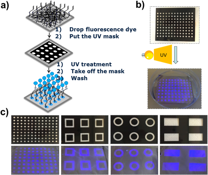
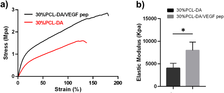
Vascular endothelial growth factor (VEGF) plays a vital role in promoting attachment and proliferation of endothelial cells, and induces angiogenesis. In recent years, much research has been conducted on functionalization of tissue engineering scaffolds with VEGF or VEGF-mimetic peptide to promote angiogenesis. However, most chemical reactions are nonspecific and require organic solvents, which can compromise control over functionalization and alter peptide/protein activity. An attractive alternative is the fabrication of functionalizable electrospun fibers, which can overcome these hurdles. In this study, we used thiol-ene chemistry for the conjugation of a VEGF-mimetic peptide to the surface of poly (ε-caprolactone) (PCL) fibrous scaffolds with varying amounts of a functional PCL-diacrylate (PCL-DA) polymer. 30% PCL-DA was selected due to homogeneous fiber morphology. A VEGF-mimetic peptide was then immobilized on PCL-DA fibrous scaffolds by a light-initiated thiol-ene reaction. 7-Mercapto-4-methylcoumarin, RGD-FITC peptide and VEGF-TAMRA mimetic peptide were used to validate the thiol-ene reaction with fibrous scaffolds. Tensile strength and elastic modulus of 30% PCL-DA fibrous scaffolds were significantly increased after the reaction. Conjugation of 30% PCL-DA fibrous scaffolds with VEGF peptide increased the surface water wettability of the scaffolds. Patterned structures could be obtained after using a photomask on the fibrous film. Moreover, in vitro studies indicated that scaffolds functionalized with the VEGF-mimetic peptide were able to induce phosphorylation of VEGF receptor and enhanced HUVECs survival, proliferation and adhesion. A chick chorioallantoic membrane (CAM) assay further indicated that the VEGF peptide functionalized scaffolds are able to promote angiogenesis in vivo. These results show that scaffold functionalization can be controlled via a simple polymer mixing approach, and that the functionalized VEGF peptide-scaffolds have potential for vascular tissue regeneration.
Keywords: Electrospun; Fibrous scaffolds; Thiol-ene reaction; VEGF peptide.
© 2022 The Authors.
Conflict of interest statement
The authors declare that they have no known competing financial interests or personal relationships that could have appeared to influence the work reported in this paper.
Figures


References
-
- Gambino L.S., Wreford N.G., Bertram J.F., Dockery P., Lederman F., Rogers P.A. Angiogenesis occurs by vessel elongation in proliferative phase human endometrium. Human Reproduction. 2002;17(5):1199–1206. - PubMed
-
- Risau W. Mechanisms of angiogenesis. nature. 1997;386(6626):671. - PubMed
-
- Laschke M.W., Harder Y., Amon M., Martin I., Farhadi J., Ring A., Torio-Padron N., Schramm R., Rücker M., Junker D. Angiogenesis in tissue engineering: breathing life into constructed tissue substitutes. Tissue engineering. 2006;12(8):2093–2104. - PubMed
LinkOut - more resources
Full Text Sources
