

Autoregulation of Cerebral Blood Flow During 3-h Continuous Cardiopulmonary Resuscitation at 27°C
- PMID: 35755426
- PMCID: PMC9218627
- DOI: 10.3389/fphys.2022.925292
Autoregulation of Cerebral Blood Flow During 3-h Continuous Cardiopulmonary Resuscitation at 27°C
Abstract
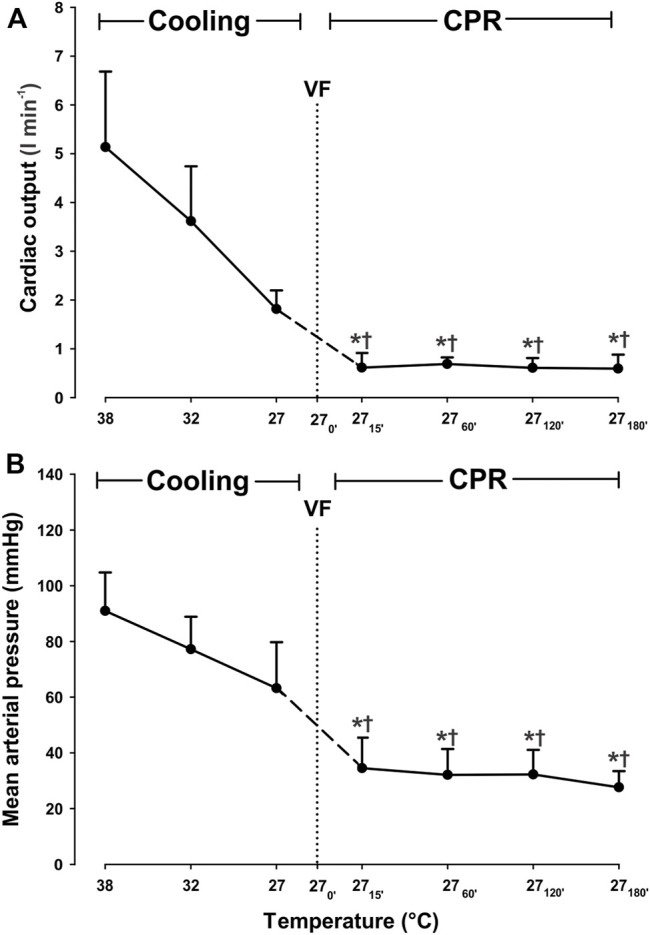
Introduction: Victims of accidental hypothermia in hypothermic cardiac arrest (HCA) may survive with favorable neurologic outcome if early and continuous prehospital cardiopulmonary resuscitation (CPR) is started and continued during evacuation and transport. The efficacy of cerebral autoregulation during hypothermic CPR is largely unknown and is aim of the present experiment. Methods: Anesthetized pigs (n = 8) were surface cooled to HCA at 27°C before 3 h continuous CPR. Central hemodynamics, cerebral O2 delivery (DO2) and uptake (VO2), cerebral blood flow (CBF), and cerebral perfusion pressure (CPP) were determined before cooling, at 32°C and at 27°C, then at 15 min after the start of CPR, and hourly thereafter. To estimate cerebral autoregulation, the static autoregulatory index (sARI), and the CBF/VO2 ratio were determined. Results: After the initial 15-min period of CPR at 27°C, cardiac output (CO) and mean arterial pressure (MAP) were reduced significantly when compared to corresponding values during spontaneous circulation at 27°C (-66.7% and -44.4%, respectively), and remained reduced during the subsequent 3-h period of CPR. During the first 2-h period of CPR at 27°C, blood flow in five different brain areas remained unchanged when compared to the level during spontaneous circulation at 27°C, but after 3 h of CPR blood flow in 2 of the 5 areas was significantly reduced. Cooling to 27°C reduced cerebral DO2 by 67.3% and VO2 by 84.4%. Cerebral VO2 was significantly reduced first after 3 h of CPR. Cerebral DO2 remained unaltered compared to corresponding levels measured during spontaneous circulation at 27°C. Cerebral autoregulation was preserved (sARI > 0.4), at least during the first 2 h of CPR. Interestingly, the CBF/VO2 ratio during spontaneous circulation at 27°C indicated the presence of an affluent cerebral DO2, whereas after CPR, the CBF/VO2 ratio returned to the level of spontaneous circulation at 38°C. Conclusion: Despite a reduced CO, continuous CPR for 3 h at 27°C provided sufficient cerebral DO2 to maintain aerobic metabolism and to preserve cerebral autoregulation during the first 2-h period of CPR. This new information supports early start and continued CPR in accidental hypothermia patients during rescue and transportation for in hospital rewarming.
Keywords: cerebral autoregulation; cerebral blood flow (CBF); cerebral oxygen consumption; cerebral oxygen delivery; cerebral perfusion pressure (CPP).
Copyright © 2022 Valkov, Nilsen, Mohyuddin, Schanche, Kondratiev, Sieck and Tveita.
Conflict of interest statement
The authors declare that the research was conducted in the absence of any commercial or financial relationships that could be construed as a potential conflict of interest.
Figures


References
LinkOut - more resources
Full Text Sources