

The Effects of Vagus Nerve Stimulation on Ventricular Electrophysiology and Nitric Oxide Release in the Rabbit Heart
- PMID: 35755432
- PMCID: PMC9213784
- DOI: 10.3389/fphys.2022.867705
The Effects of Vagus Nerve Stimulation on Ventricular Electrophysiology and Nitric Oxide Release in the Rabbit Heart
Abstract
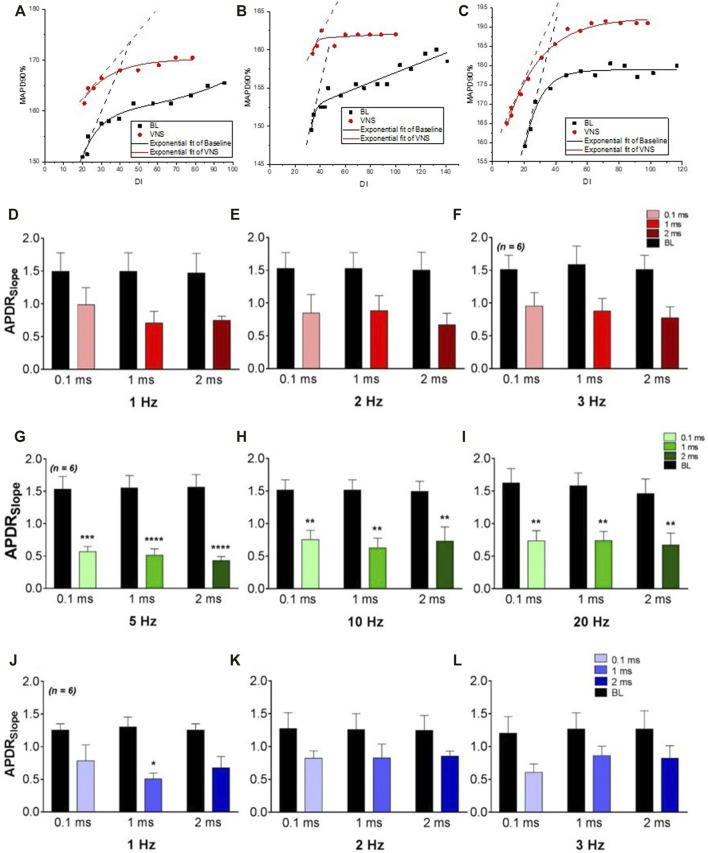
Background: Abnormal autonomic activity including impaired parasympathetic control is a known hallmark of heart failure (HF). Vagus nerve stimulation (VNS) has been shown to reduce the susceptibility of the heart to ventricular fibrillation, however the precise underlying mechanisms are not well understood and the detailed stimulation parameters needed to improve patient outcomes clinically are currently inconclusive. Objective: To investigate NO release and cardiac electrophysiological effects of electrical stimulation of the vagus nerve at varying parameters using the isolated innervated rabbit heart preparation. Methods: The right cervical vagus nerve was electrically stimulated in the innervated isolated rabbit heart preparation (n = 30). Heart rate (HR), effective refractory period (ERP), ventricular fibrillation threshold (VFT) and electrical restitution were measured as well as NO release from the left ventricle. Results: High voltage with low frequency VNS resulted in the most significant reduction in HR (by -20.6 ± 3.3%, -25.7 ± 3.0% and -30.5 ± 3.0% at 0.1, 1 and 2 ms pulse widths, with minimal increase in NO release. Low voltage and high frequency VNS significantly altered NO release in the left ventricle, whilst significantly flattening the slope of restitution and significantly increasing VFT. HR changes however using low voltage, high frequency VNS were minimal at 20Hz (to 138.5 ± 7.7 bpm (-7.3 ± 2.0%) at 1 ms pulse width and 141.1 ± 6.6 bpm (-4.4 ± 1.1%) at 2 ms pulse width). Conclusion: The protective effects of the VNS are independent of HR reductions demonstrating the likelihood of such effects being as a result of the modulation of more than one molecular pathway. Altering the parameters of VNS impacts neural fibre recruitment in the ventricle; influencing changes in ventricular electrophysiology, the protective effect of VNS against VF and the release of NO from the left ventricle.
Keywords: autonomic nervous system; electrophysiology; heart; nitric oxide; vagus nerve stimulation.
Copyright © 2022 Allen, Pongpaopattanakul, Chauhan, Brack and Ng.
Conflict of interest statement
The authors declare that the research was conducted in the absence of any commercial or financial relationships that could be construed as a potential conflict of interest.
Figures


References
-
- Allen E., Coote J. H., Grubb B. D., Batten T. F. C., Pauza D. H., Ng G. A., et al. (2018). Electrophysiological Effects of Nicotinic and Electrical Stimulation of Intrinsic Cardiac Ganglia in the Absence of Extrinsic Autonomic Nerves in the Rabbit Heart. Heart Rhythm 15, 1698–1707. 10.1016/j.hrthm.2018.05.018 - DOI - PMC - PubMed
Grants and funding
LinkOut - more resources
Full Text Sources
Research Materials
Miscellaneous