Review
. 2022 May;10(5):366-380.
doi: 10.22038/ABJS.2021.60054.2958.

Meniscal Root Tears: A Decade of Research on their Relevant Anatomy, Biomechanics, Diagnosis, and Treatment

Affiliations
Review

Meniscal Root Tears: A Decade of Research on their Relevant Anatomy, Biomechanics, Diagnosis, and Treatment

Mark T Banovetz et al. Arch Bone Jt Surg. 2022 May.

Abstract

A foundational knowledge of the anatomy and biomechanics of meniscal root tears is warranted for proper repair of meniscal root tears and for preventing some of their commonly described iatrogenic causes. Meniscal root tears are defined as either a radial tear occurring within one cm of the root attachment site of the meniscus or a complete bony or soft tissue avulsion of the root attachment altogether. Meniscal root tears disrupt the protective biomechanical function of the native meniscus. Biomechanical analyses of the current techniques for meniscal root repair highlight the importance of restoring menisci to their correct anatomic orientation, thereby restoring their biomechanical function. A comprehensive understanding of the clinical and radiographic presentations of these injuries is critical to preventing their underdiagnosis. The poor long-term outcomes associated with conservative treatment measures, namely, ipsilateral compartment osteoarthritis, warrants the surgical repair of meniscal root tears whenever possible. While excellent patient-reported outcomes exist for the various surgical repair techniques, adherence to stringent post-operative rehabilitation protocols is critical for patients to avoid damaging the integrity of a repaired root. This review will focus on current concepts pertaining to the anatomy, biomechanics, diagnosis, treatment, and postoperative rehabilitation for meniscal root tears.

Keywords: Anterior cruciate ligament; Meniscus; Root.


Figures

Figure 1
Figure 1
Photograph of a superoposterior view of the tibial plateau demonstrating the qualitative anatomic relationships between important fibrocartilaginous structures and ligamentous insertions. MM, medial meniscus; LM, lateral meniscus; B, body; AH, anterior horn; PH, posterior horn; Rt, root; SWFs, shiny white fibers; AIL, anterior intermeniscal ligament
Figure 2
Figure 2
Photograph of a superoanterior view of the tibial plateau with lines superimposed on the medial meniscus approximating the boundaries of the three vascular zones of the meniscus. WW, white-white zone; RW, red-white zone; RR, red-red zone
Figure 3
Figure 3
Illustration of the tibial plateau from a superior view (A) and posterior view (B). The quantitative anatomical dimensions of the LPRA and MPRA are superimposed upon (A). In both (A) and (B), the footprints of the tibial insertions for both the ACL and PCL are depicted as darkened areas in their respective locations. The shiny white fibers of the MPRA are denoted SWF in both (A) and (B). Reproduced with permission from AJSM Vol. 40 Issue 10, 2342-2347
Figure 4
Figure 4
Photographs depicting the tibial plateau of a human cadaveric dissection from a superior view (A) and posterior view (B). The MPRA and LPRA are shown intact in (A) and severed in (B). The native locations of the MPRA and LPRA are approximated with the blue outlining in (B). Reproduced with permission from AJSM Vol. 40 Issue 10, 2342-2347
Figure 5
Figure 5
Illustration created by LaPrade et al. depicting the tibial plateau structures and emphasizing the overlap of the LARA and the tibial insertion of the ACL. The authors created a “danger zone” for repair of the ACL using the quantitative anatomical overlap between the tibial insertions for the two structures, with colors approaching the color red along the provided spectrum indicating a higher frequency of insertion in the cadaveric specimens examined by the authors. SFs, shiny white fibers of the MARA; AM root, MARA center; ACL center, center of the tibial insertion of the ACL; AL root, LARA center, AC, articular cartilage; LTE Apex, apex of the lateral tibial eminence; PL, LPRA center; PM, MPRA center; TT, tibial tubercle. Reproduced with permission from AJSM Vol. 42 Issue 10, pp. 2386-2392
Figure 6
Figure 6
Series of photographs depicting a torn meniscal root belonging to a young, healthy patient on arthroscopy, as well its subsequent repair. (A) a probe is used to place superiorly directed tension on the root attachment site. Because the root is completely torn, the torn root is elevated off of the tibial plateau. (B) a guide is placed over the native root attachment site of the tibial plateau in order to guide drilling. (C) a guide pin is drilled through the native root attachment site on the tibial plateau. (D) the meniscal root is secured to its native attachment site by two sutures situated anteroposterior to each other
Figure 7
Figure 7
Photograph demonstrating palpation of the medial joint line of the knee in order to check for the presence of palpable meniscal extrusion
Figure 8
Figure 8
Photographs taken in succession, demonstrating the pivot shift maneuver performed under general anesthesia in the context of a patient with ACL deficiency and concomitant meniscal root pathology. Here, the patient presents with a 3+ “explosive” finding on the pivot shift test. (A) depicts the clinician applying a superomedial force on the leg. (B) demonstrates subluxation of the proximal tibia against the distal femur. (C) is taken immediately following the “explosion” as the tibia is reduced out of its originally subluxated state by the IT band. (D) depicts a reversal of the direction of the force applied by the clinician and is taken immediately following the point where the tibia has returned to its subluxated position
Figure 9
Figure 9
Illustration depicting the five types of meniscal root tears as classified by LaPrade et al. All of the tears are depicted as occurring at the MPRA. Reproduced with permission from AJSM Vol. 43 Issue 2, pp. 363-369
Figure 10
Figure 10
Photograph depicting the probing of a posterior meniscal root on arthroscopy. The meniscal root depicted here was previously repaired, and this photograph demonstrates that it is intact
Figure 11
Figure 11
Photographs depicting the five types of meniscal root tears visualized arthroscopically. (A) depicts a type 1 meniscal root tear. (B) depicts a type 2 meniscal root tear. (C) depicts a type 3 meniscal root tear. (D) depicts a type 4 meniscal root tear. (E) depicts a type 5 meniscal root tear. Arrows mark the location of the tear depicted in each photograph. F, femur; T, tibia; RT, root tear; BHT, bucket-handle tear; AV, avulsion. Reproduced with permission from AJSM Vol. 43 Issue 2, pp. 363-369
Figure 12
Figure 12
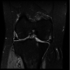
Coronal MRI radiographs depicting extrusion of the (A) medial, and (B) lateral, meniscus following a meniscal root tear. (A) depicts an MRI radiograph of a left knee with visible medial extrusion of the medial meniscus. (B) depicts an MRI radiograph of a right knee with visible lateral extrusion of the lateral meniscus. The extrusion depicted in both (A) and (B) exceeds the threshold of 3 mm between the outer margin of the tibial plateau articular cartilage and the outer extent of the meniscus
Figure 13
Figure 13
(A) coronal and (B) sagittal MRI radiographs depicting spontaneous osteonecrosis of the knee (SONK)/Subchondral insufficiency fracture (SIFK) of the medial femoral condyle following medial meniscal root tear. Significant medial meniscal extrusion is also present in (A).
Figure 14
Figure 14
Sagittal MRI radiographs depicting (A) a normal cross-sectional view of the meniscus versus visible ghost sign of the meniscus in the same sequence (B), and in a different sequence (C).
Figure 15
Figure 15
Coronal MRI radiograph following the surgical repair of a meniscal root tear demonstrating persistent meniscal extrusion. The extrusion depicted here is observably significant, measuring greater than 3 mm. This repair underwent subsequent revision
Figure 16
Figure 16
Illustration depicting the repair of a tear to the MPRA using a transtibial pull-out technique. The specific technique depicted above uses two transosseous tunnels and two sutures tied over a cortical button. The sutures are passed through the meniscal root anteroposterior to each other. Reproduced with permission from AJSM Vol. 45 Issue 4, 884-891
Figure 17
Figure 17
Photographs taken on arthroscopy during a transtibial repair of a meniscal root tear. (A) photograph taken before peripheral release of the meniscus has been performed. Peripheral adhesion of the meniscus to the joint capsule has caused apparent subluxation of the meniscus, complicating anatomic reduction of the meniscal root to its native attachment site for repair. (B) photograph taken subsequent to peripheral release of the meniscus from the capsule. Now free of adhesion to the capsule, the meniscus is available to occupy a visibly greater area greater of the joint space and assume a more native orientation, making successful anatomic reduction and fixation of the meniscal root possible
Figure 18
Figure 18
Images taken on arthroscopy depicting a complete tear of the LPRA (A) and subsequent anatomic reapproximation and fixation using two sutures, which have been pulled through transtibial tunnels and secured over the proximal tibial cortex using a cortical button. Reproduced with permission from AJSM Vol. 45 Issue 4, 884-891

Similar articles

Cited by

References

    1. LaPrade RF, Floyd ER, Carlson GB, Moatshe G, Chahla J, Monson JK. Meniscal root tears: Solving the silent epidemic. Journal of Arthroscopic Surgery and Sports Medicine. 2021;2(1):47–57.
    1. LaPrade CM, Jansson KS, Dornan G, Smith SD, Wijdicks CA, LaPrade RF. Altered tibiofemoral contact mechanics due to lateral meniscus posterior horn root avulsions and radial tears can be restored with in situ pull-out suture repairs. J Bone Joint Surg Am. 2014;96(6):471–9. - PubMed
    1. Steineman BD, LaPrade RF, Santangelo KS, Warner BT, Goodrich LR, Haut Donahue TL. Early Osteoarthritis After Untreated Anterior Meniscal Root Tears: An In Vivo Animal Study. Orthop J Sports Med. 2017;5(4):2325967117702452. - PMC - PubMed
    1. Cinque ME, DePhillipo NN, Moatshe G, Chahla J, Kennedy MI, Dornan GJ, et al. Clinical Outcomes of Inside-Out Meniscal Repair According to Anatomic Zone of the Meniscal Tear. Orthop J Sports Med. 2019;7(7):2325967119860806. - PMC - PubMed
    1. Matheny LM, Ockuly AC, Steadman JR, LaPrade RF. Posterior meniscus root tears: associated pathologies to assist as diagnostic tools. Knee Surg Sports Traumatol Arthrosc. 2015;23(10):3127–31. - PubMed

LinkOut - more resources