. 2022 Jun 8:9:896493.
doi: 10.3389/fmolb.2022.896493. eCollection 2022.

Multivalent Interaction of Beta-Catenin With its Intrinsically Disordered Binding Partner Adenomatous Polyposis Coli

Affiliations

Multivalent Interaction of Beta-Catenin With its Intrinsically Disordered Binding Partner Adenomatous Polyposis Coli

Pamela J E Rowling et al. Front Mol Biosci. .

Abstract

The Wnt signalling pathway plays key roles in cell proliferation, differentiation and fate decisions in embryonic development and maintenance of adult tissues, and the twelve Armadillo (ARM) repeat-containing protein β-catenin acts as the signal transducer in this pathway. Here we investigate the interaction between β-catenin's ARM repeat domain and the intrinsically disordered protein adenomatous polyposis coli (APC). APC is a giant multivalent scaffold that brings together the different components of the so-called "β-catenin destruction complex", which drives β-catenin degradation via the ubiquitin-proteasome pathway. Mutations and truncations in APC, resulting in loss of APC function and hence elevated β-catenin levels and upregulation of Wnt signalling, are associated with numerous cancers including colorectal carcinomas. APC has a long intrinsically disordered region (IDR) that contains a series of 15-residue and 20-residue binding regions for β-catenin. Here we explore the multivalent nature of the interaction of β-catenin with the highest affinity APC repeat, both at equilibrium and under kinetic conditions. We use a combination of single-site substitutions, deletions and insertions to dissect the mechanism of molecular recognition and the roles of the three β-catenin-binding subdomains of APC.

Keywords: adenomatous polyposis coli (APC); armadillo repeat; beta-catenin (β-catenin); fuzzy binding; intrinsically disordered protein; multivalency; protein-protein interaction (PPI).


Conflict of interest statement

The authors declare that the research was conducted in the absence of any commercial or financial relationships that could be construed as a potential conflict of interest.

Figures

FIGURE 1
FIGURE 1
APC and β-catenin. (A) Schematic of the domain structure of APC from N- to C-terminus: coiled-coil oligomerisation domain, turquoise; armadillo domain, light green; β-catenin- binding 15aa repeats, red; β-catenin-binding 20aa repeats, pink; axin-binding SAMP repeats, yellow; microtubule-binding basic region, blue; and EB1-binding domain, purple. The mutational cluster region is underlined in red. (B) Sequence of the third 20aa repeat construct of APC, and fragments thereof, used in this study. Phosphorylated residues are in bold, and lysine-interacting residues are underlined. (C) Prediction of disorder propensity of the third 20aa repeat (R3) of APC using the flDPnn program. Amino acids with values above the dotted line are predicted to be disordered and those below ordered (Hu et al., 2021). The coloured bars at the top of the graph represent the three subdomains of APC R3: the N-terminal α-helical domain is in peach (APCa), the lysine-binding domain in purple (APCb), and the phosphorylation domain in pink (APCc).
FIGURE 2
FIGURE 2
The interaction of the third 20aa repeat (R3) of APC with β-catenin. (A) Schematic representation of the structure of the armadillo repeat domain of β-catenin in complex with the phosphorylated third 20aa repeat of APC(R3) (PDB 1TH1) (Xing et al., 2004). The three subdomains of APC R3 are coloured: the N-terminal α-helical domain in peach (APCa), the lysine binding domain in purple (APCb), and the phosphorylation domain in pink (APCc). The amino acids that are phosphorylated are represented as sticks and the two lysine-interacting amino acids as spheres. The β-catenin ARM repeats that bind to the three APC subdomains are coloured: ARM 10–12 in blue, ARM 5–9 in turquoise, and ARM 1–4 in dark blue. (B) Schematic showing the location of the key β-catenin-binding residues in APC R3. APC and β-catenin are coloured as in (A). The five phosphorylated residues, S1–S4 and T1, are represented by circles. The lysine-binding residues D1486 and E1494 are represented by triangles. The site of the single cysteine residue (C1501) is represented by a yellow star. (C) Buried surface area by residue of pAPC R3 in the complex with β-catenin derived from PDB 1TH1 calculated using Cocomaps (http://www.molnac.unisa.it/BioTools/cocomaps, Vangone et al., 2011). The bars at the top of the graph indicate the three subdomains of APC R3 coloured as in (A).
FIGURE 3
FIGURE 3
Increasing affinity for β-catenin of APCbc constructs with increasing number of phosphomimetic substitutions. Glutamate substitutions were made at phosphorylation sites T1487E (T1), S1504E (S1), S1505E (S2), S1507E (S3), S1510E (S4). The IC50s of these phosphomimetic APC bc variants was measured by fluorescence competition assay using APC bc labelled with Alexa488 at Cys 1501. The data are shown as the mean ± SEM of three replicates.
FIGURE 4
FIGURE 4
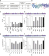
Effects of truncations and mutations in APC on β-catenin-binding. Dissociation constants (A), on-rates (B), off-rates (C), and time constants (D). Both fast and slow off-rates are shown. The time constant are calculated from the fast off-rates.
FIGURE 5
FIGURE 5
Binding of β-catenin to the unphosphorylated and phosphomimetic forms of APCabc. The dissociation constants were measured by fluorescence anisotropy using APCabc labelled with fluorescein at Cys 1501. The unphosphorylated APCabc is shown in black circles and the phosphomimetic, APCabc, in open circles. The data are shown as the mean ± SEM of at least three replicates.
FIGURE 6
FIGURE 6
The effect of linker insertions and mutation of the lysine-interaction residue D1486S in APC on β-catenin-binding. (A) The sequences and properties of the linkers inserted into APCabc. The hydropathy was calculated using mean hydropathy of a sequence (Kyte and Doolittle, 1982). (B) Schematic showing the two sites of linker insertions in APC. (C) Dissociation constants were measured by fluorescence anisotropy of the APC variants labelled with fluorescein at Cys 1501, and the data are shown as the mean ± SEM of three replicates. (D) The association rates and the rates of the two dissociation phases, fast (E) and slow (F), were measured by stopped-flow fluorescence. The data shown are the mean ± SEM of at least two replicate experiments consisting of five kinetic traces at each concentration measured.
FIGURE 7
FIGURE 7
Schematic of the contributions of the different subdomains of APC to the interaction with β-catenin. The key contacting residues are denoted in triangles (lysine-binding residues) and circles (phosphomimetics). Key dissociation constants and rates of association and dissociation are shown. The intrinsic β-catenin-binding affinity of the b domain can be estimated from the affinity of APC, which has a weakened c domain. The intrinsic β-catenin-binding affinity of the c domain can be estimated from the affinity of pAPC D1486S, which has a weakened b domain.

Similar articles

Cited by

References

    1. Adzhubei A. A., Sternberg M. J. E., Makarov A. A. (2013). Polyproline-II Helix in Proteins: Structure and Function. J. Mol. Biol. 425, 2100–2132. 10.1016/j.jmb.2013.03.018 - DOI - PubMed
    1. Aksel T., Barrick D. (2014). Direct Observation of Parallel Folding Pathways Revealed Using a Symmetric Repeat Protein System. Biophysical J. 107, 220–232. 10.1016/j.bpj.2014.04.058 - DOI - PMC - PubMed
    1. Arai R. (2021). Design of Helical Linkers for Fusion Proteins and Protein-Based Nanostructures. Methods Enzymol. 647, 209–230. 10.1016/BS.MIE.2020.10.003 - DOI - PubMed
    1. Bachmann A., Wildemann D., Praetorius F., Fischer G., Kiefhaber T. (2011). Mapping Backbone and Side-Chain Interactionsin the Transition State of a Coupled Proteinfolding and Binding Reaction. Proc. Natl. Acad. Sci. U. S. A. 104, 3952–3957. 10.1073/pnas.1012668108 - DOI - PMC - PubMed
    1. Borg M., Mittag T., Pawson T., Tyers M., Forman-Kay J. D., Chan H. S. (2007). Polyelectrostatic Interactions of Disordered Ligands Suggest a Physical Basis for Ultrasensitivity. Proc. Natl. Acad. Sci. U.S.A. 104, 9650–9655. 10.1073/PNAS.0702580104 - DOI - PMC - PubMed

LinkOut - more resources