. 2022 Jun 9:13:883650.
doi: 10.3389/fmicb.2022.883650. eCollection 2022.

Gut Microbiota Characteristics Are Associated With Severity of Acute Radiation-Induced Esophagitis

Affiliations

Gut Microbiota Characteristics Are Associated With Severity of Acute Radiation-Induced Esophagitis

Ming-Qiang Lin et al. Front Microbiol. .

Abstract

Background: Acute radiation-induced esophagitis (ARIE) is one of the most debilitating complications in patients who receive thoracic radiotherapy, especially those with esophageal cancer (EC). There is little known about the impact of the characteristics of gut microbiota on the initiation and severity of ARIE.

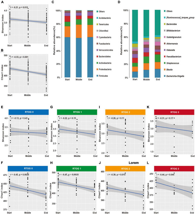
Materials and methods: Gut microbiota samples of EC patients undergoing radiotherapy (n = 7) or concurrent chemoradiotherapy (n = 42) were collected at the start, middle, and end of the radiotherapy regimen. Assessment of patient-reported ARIE was also performed. Based on 16S rRNA gene sequencing, changes of the gut microbial community during the treatment regimen and correlations of the gut microbiota characteristics with the severity of ARIE were investigated.

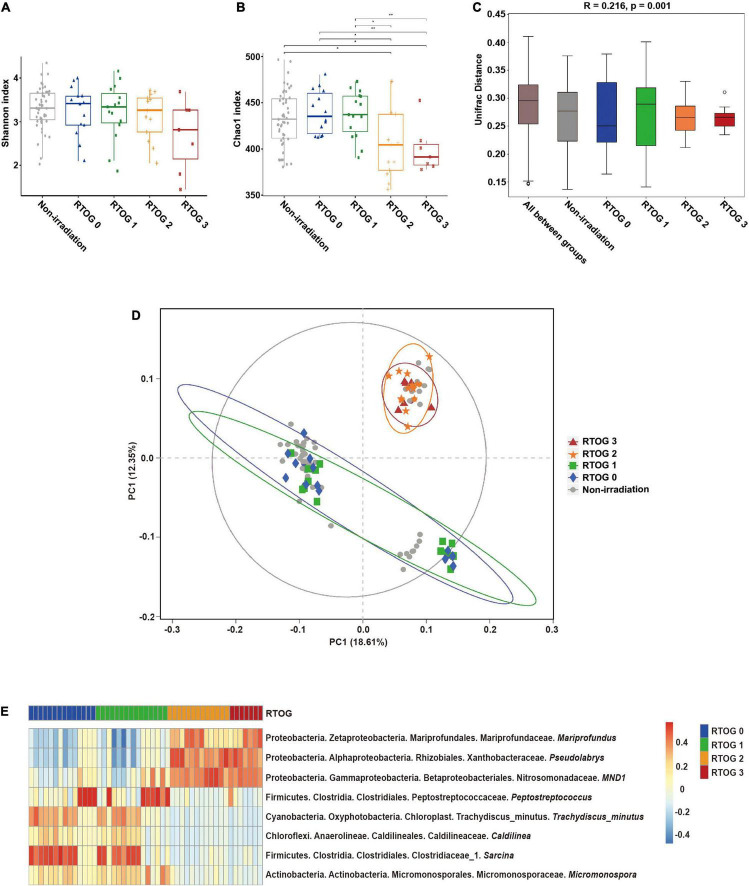
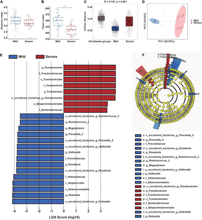
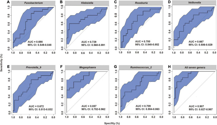
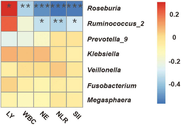
Results: There were significant associations of several properties of the gut microbiota with the severity of ARIE. The relative abundance of several genera in the phylum Proteobacteria increased significantly as mucositis severity increased. The predominant genera had characteristic changes during the treatment regimen, such as an increase of opportunistic pathogenic bacteria including Streptococcus. Patients with severe ARIE had significantly lower alpha diversity and a higher abundance of Fusobacterium before radiotherapy, but patients with mild ARIE were enriched in Klebsiella, Roseburia, Veillonella, Prevotella_9, Megasphaera, and Ruminococcus_2. A model combining these genera had the best performance in prediction of severe ARIE (area under the curve: 0.907).

Conclusion: The characteristics of gut microbiota before radiotherapy were associated with subsequent ARIE severity. Microbiota-based strategies have potential use for the early prediction of subsequent ARIE and for the selection of interventions that may prevent severe ARIE.

Keywords: 16S rRNA gene; acute radiation-induced esophagitis; esophageal cancer; gut microbiota; radiotherapy.


Conflict of interest statement

The authors declare that the research was conducted in the absence of any commercial or financial relationships that could be construed as a potential conflict of interest.

Figures

FIGURE 1
FIGURE 1
Relationship of ARIE severity with fecal microbial community structure of samples collected at “middle” of the radiotherapy regimen. (A,B) Alpha diversity indexes (Shannon and Chao1) of the non-irradiation (gray), RTOG 0 (blue), RTOG 1 (green), RTOG 2 (orange), and RTOG 3 (red) groups. A larger index indicates greater community diversity. (C) Box plot of inter-group and intra-group Unifrac distance-based ANOSIM. R-value > 0 indicated that the inter-group difference was greater than the intra-group difference. (D) PCoA of microbial community structure in the non-irradiation group and each RTOG group. Each sample is represented by a symbol, and symbols with different colors and shapes correspond to different groups. (E) Spearman correlation heatmap of the relationship of ARIE severity (RTOG grade) with the relative abundance of eight bacterial genera (| r| ≥ 0.5, p < 0.05), in which the relative abundance of each genus was converted to a log10 value, indicated by a color gradient. *P < 0.05; **P < 0.01.
FIGURE 2
FIGURE 2
Changes in fecal microbial community structure during chemoradiotherapy. (A,B) Changes in alpha-diversity indexes (Shannon and Chao1) in the overall microbial community. (C,D) Average relative abundances of predominant bacterial taxa at the phylum and genus levels. Changes of alpha diversity indexes (Shannon and Chao1) in the RTOG 0 (E,F), RTOG 1 (G,H), RTOG 2 (I,J), and RTOG 3 (K,L) groups during the course of chemoradiotherapy.
FIGURE 3
FIGURE 3
Gut microbial community structure before radiotherapy (“start”) in patients who subsequently experienced mild or severe ARIE. (A,B) Alpha-diversity indexes (Shannon and Chao1) of the mild (blue) and severe (red) subgroups. (C) Box plot of inter-group and intra-group Unifrac distance-based ANOSIM. R-value > 0 indicated the inter-group difference was greater than the intra-group difference. (D) PCoA of Unifrac distance of the mild and severe subgroups. (E,F) LEfSe analysis (LDA significance threshold > 3.5) of taxa with the greatest differences in abundance between the severe (red, positive scores) and mild (blue, negative scores) subgroups. ****P < 0.0001.
FIGURE 4
FIGURE 4
Receiver operating characteristic (ROC) analysis of the predictive value of the abundance of seven fecal microbial genera prior to radiotherapy on subsequent severity of ARIE. (A) Fusobacterium, (B) Klebsiella, (C) Roseburia, (D) Veillonella, (E) Prevotella_9, (F) Megasphaera, (G) Ruminococcus_2, (H) All seven genera.
FIGURE 5
FIGURE 5
Spearman correlation heatmap between fecal microbial genera and clinical indicators of immune-inflammation before radiotherapy (“start”). WBC, white blood cells; NE, neutrophils; LY, lymphocytes; NLR, neutrophil-lymphocyte ratio; SII, systemic immune inflammation index (platelets × NLR). *P < 0.05; **P < 0.01; ***P < 0.001.

References

    1. Adebahr S., Schimek-Jasch T., Nestle U., Brunner T. B. (2016). Oesophagus side effects related to the treatment of oesophageal cancer or radiotherapy of other thoracic malignancies. Best Pract. Res. Clin. Gastroenterol. 30 565–580. 10.1016/j.bpg.2016.07.003 - DOI - PubMed
    1. Ajani J. A., D’Amico T. A., Bentrem D. J., Chao J., Corvera C., Das P., et al. (2019). Esophageal and esophagogastric junction cancers, version 2.2019, NCCN clinical practice guidelines in oncology. J. Natl. Compr. Cancer Netw. 17 855–883. 10.6004/jnccn.2019.0033 - DOI - PubMed
    1. Albillos A., De Gottardi A., Rescigno M. (2020). The gut-liver axis in liver disease: pathophysiological basis for therapy. J. Hepatol. 72 558–577. 10.1016/j.jhep.2019.10.003 - DOI - PubMed
    1. Bolger A. M., Lohse M., Usadel B. (2014). Trimmomatic: a flexible trimmer for Illumina sequence data. Bioinformatics 30 2114–2120. 10.1093/bioinformatics/btu170 - DOI - PMC - PubMed
    1. Bradley J., Movsas B. (2004). Radiation esophagitis: predictive factors and preventive strategies. Semin. Radiat. Oncol. 14 280–286. 10.1016/j.semradonc.2004.06.003 - DOI - PubMed

LinkOut - more resources