. 2022 Jun 10:13:872505.
doi: 10.3389/fmicb.2022.872505. eCollection 2022.

Sophoridine Suppresses Herpes Simplex Virus Type 1 Infection by Blocking the Activation of Cellular PI3K/Akt and p38 MAPK Pathways

Affiliations

Sophoridine Suppresses Herpes Simplex Virus Type 1 Infection by Blocking the Activation of Cellular PI3K/Akt and p38 MAPK Pathways

Qiong Tang et al. Front Microbiol. .

Abstract

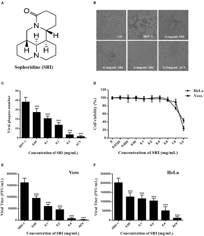
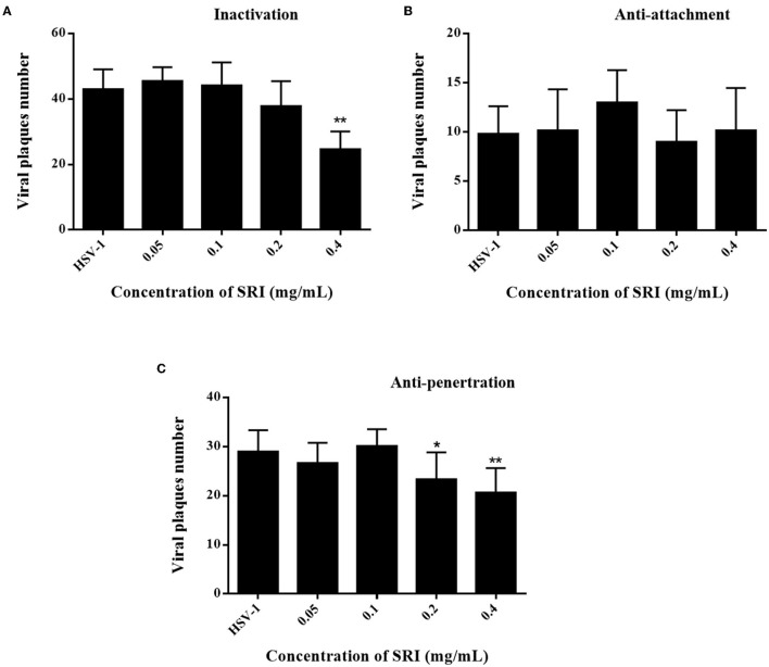
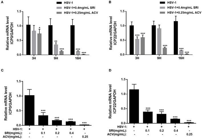
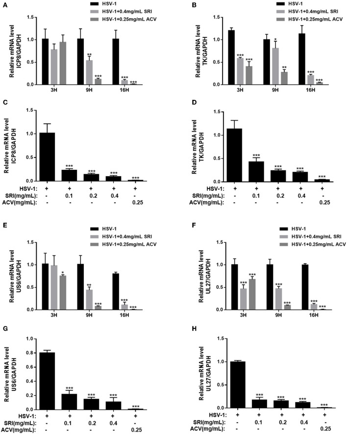
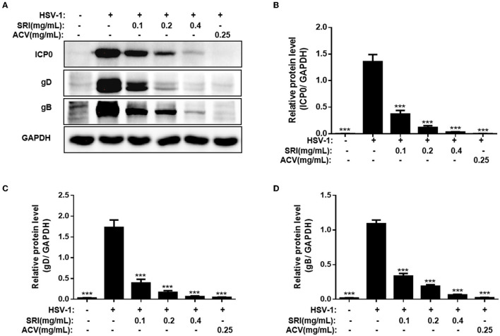
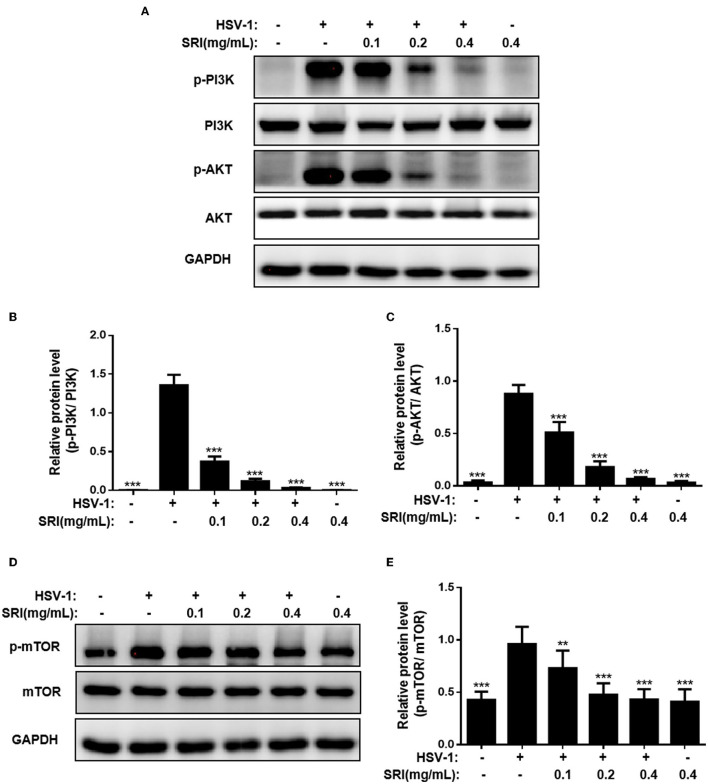
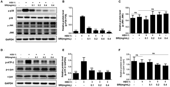
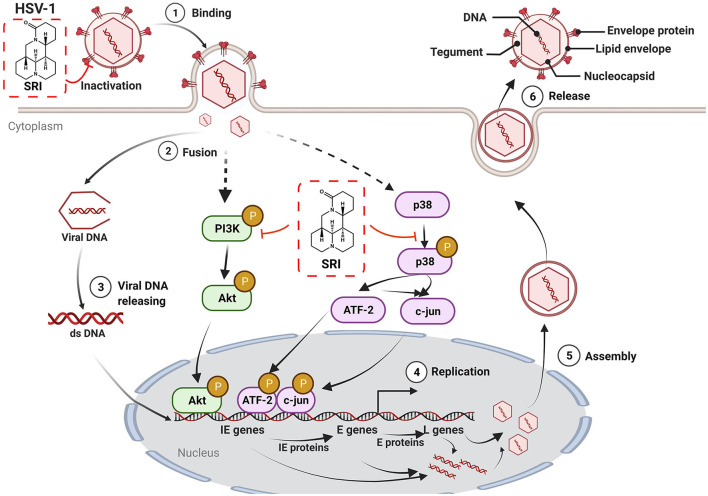
Herpes simplex virus type 1 (HSV-1) is a ubiquitous and important human pathogen capable of causing significant clinical diseases ranging from skin damage to encephalitis, particularly in immunocompromised and neonatal hosts. Currently, widely used nucleoside analogs, including acyclovir and penciclovir, have some limitations in their use due to side effects and drug resistance. Herein, we report sophoridine's (SRI) dramatic inhibition of HSV-1 replication in vitro. SRI exhibited a remarkable inhibitory influence on HSV-1 virus-induced cytopathic effect and plaque formation, as well as on progeny viruses in Vero and HeLa cells, with selection indexes (SI) of 38.96 and 22.62, respectively. Moreover, SRI also considerably suppressed HSV-1 replication by hindering the expression of viral immediate-early (ICP0 and ICP22), early (ICP8 and TK), and late (gB and gD) genes and the expression of viral proteins ICP0, gB, and gD. We suggest that SRI can directly inactivate viral particles and block some stages in the life cycle of HSV-1 after adsorption. Further experiments showed that SRI downregulated the cellular PI3K/Akt signaling pathway and obstructed HSV-1 replication even more. Most importantly, SRI markedly repressed HSV-1-induced p38 MAPK pathway activation. Collectively, this natural bioactive alkaloid could be a promising therapeutic candidate against HSV-1 via the modulation of cellular PI3K/Akt and p38 MAPK pathways.

Keywords: HSV-1; PI3K/Akt pathway; antiviral; p38 MAPK pathway; sophoridine.


Conflict of interest statement

The authors declare that the research was conducted in the absence of any commercial or financial relationships that could be construed as a potential conflict of interest.

Figures

Figure 1
Figure 1
Toxicities and inhibitory effects of SRI on HSV-1 in vitro. (A) The chemical structure of SRI. (B) The cells infected with HSV-1 (MOI = 1) were treated with SRI (0.1, 0.2, and 0.4 mg/mL) or ACV (0.25 mg/mL), and morphological changes of Vero cells were captured under an optical inverted microscope at 72 h post-infection. (C) The cells infected with HSV-1 (MOI = 1) were treated with SRI or ACV, and the inhibitory effect of SRI was estimated by plaque assay. The number of plaques on Vero cells infected with HSV-1 was counted. (D) Vero and HeLa cells were treated with SRI (from 0.0125 to 3.2 mg/mL) for 48 or 72 h, and cell viability was calculated by MTT assay. (E,F) Vero and HeLa cells were infected with HSV-1 (MOI = 1) and were then treated with SRI or ACV for 24 h. The progeny virus was released after three cycles of freezing and thawing of the infected cells, and the progeny virus titer was determined by plaque assay. The results are given as mean ± SD. ***p < 0.001 vs. HSV-1 group.
Figure 2
Figure 2
Effects of different treatment conditions of SRI on HSV-1 infection. (A) Pre-treatment of virus: HSV-1 was pre-treated with SRI (0.05, 0.1, 0.2, and 0.4 mg/mL) and incubated at 37°C for 1 h. The viral inoculum was added to a Vero cell monolayer and incubated for 2 h. Then, a plaque reduction assay was performed to calculate the number of plaques. (B) Viral attachment: pre-chilled monolayer of Vero cells was infected with HVS-1 and SRI (0.05, 0.1, 0.2, and 0.4 mg/mL) and incubated for 2 h at 4°C to allow binding (but not cellular uptake). Then, a plaque reduction assay was performed to calculate the number of plaques. (C) Viral penetration: HSV-1 was added to a pre-chilled monolayer of Vero cells. After incubation at 4°C for 2 h, the viruses were removed and Vero cells were incubated with SRI (0.05, 0.1, 0.2, and 0.4 mg/mL) at 37°C for 2 h to facilitate viral penetration. Then plaque reduction assay was performed to calculate the number of plaques. The results are presented as mean ± SD. *p < 0.05, **p < 0.01 vs. HSV-1 group.
Figure 3
Figure 3
SRI repressed HSV-1 IE gene expression. (A–D) Confluent cells were infected with HSV-1 for 2 h and then treated with the designated concentrations of SRI (0.05, 0.1, 0.2, and 0.4 mg/mL) or ACV (0.25 mg/mL). Total RNA was extracted at the indicated time points (3, 9, and 16 h) and RT-PCR was employed to analyze the ICP0 and ICP22 mRNA levels (A,B). The effects of SRI and ACV on ICP0 and ICP22 genes of the virus were determined after 16 h (C,D). The level of gene transcription was normalized by GAPDH. The data are presented as mean ± SD. *p < 0.05, **p < 0.01, and ***p < 0.001, vs. HSV-1 group.
Figure 4
Figure 4
Effect of SRI on HSV-1 E and L gene expression. (A–H) Confluent cells were infected with HSV-1 for 2 h and then treated with the designated concentrations of SRI (0.05, 0.1, 0.2, and 0.4 mg/mL) or ACV (0.25 mg/mL). Total RNA was extracted at the indicated time points (3, 9, and 16 h) and RT-PCR was employed to analyze the ICP8 (A), TK (B), US6 (E), and UL27 (F) mRNA levels. The effects of SRI and ACV on ICP8, TK, US6, and UL27 genes of virus were determined after 16 h (C,D,G,H). The level of gene transcription was normalized by GAPDH. The data were presented as mean ± SD. *p < 0.05, **p < 0.01, and ***p < 0.001, vs. HSV-1 group.
Figure 5
Figure 5
SRI inhibited the HSV-1 ICP0, gB, and gD protein expression levels. (A–D) Cells grown in six-well plate were infected with HSV-1 and treated with SRI (0.05, 0.1, 0.2, and 0.4 mg/mL) or ACV (0.25 mg/mL). At 24 h post-infection, the cellular proteins were harvested and determined using Western blot analysis using primary antibodies to HSV-1 ICP0, gB, gD, and GAPDH. GAPDH was used as a standard loading control. The quantification results of the Western blot analysis of ICP0 (B), gD (C), and gB (D). The data are expressed as mean ± SD. ***p < 0.001, vs. HSV-1 group.
Figure 6
Figure 6
SRI inhibited HSV-1 replication through blocking of PI3K/Akt pathway. (A–E) The infected cells of HSV-1 (MOI = 1) were treated with or without SRI (0.1, 0.2, and 0.4 mg/mL), and the protein levels of p-PI3K, PI3K, p-Akt, and Akt were evaluated using Western blotting analysis. Blots were also probed for GAPDH, which was used as a loading control. Plots quantifying the immunoblots as ratios for p-PI3K/PI3K (B), p-AKT/Akt (C), and p-mTOR/mTOR (E), respectively. The results shown are representative of three independent experiments. The results are presented as mean ± SD values. **p < 0.01 and ***p < 0.001 vs HSV-1 group; ns, not significant.
Figure 7
Figure 7
SRI restrained HSV-1 replication through blocking of P38 MAPK pathway. (A,D) The infected cells of HSV-1 (MOI = 1) were treated without or with SRI (0.1, 0.2, and 0.4 mg/mL), and then the protein levels of p-p38, p38, JNK, p-JNK, p-ATF-2, p-c-jun, and c-jun were evaluated through Western blot analysis. Blots were also probed for GAPDH and tubulin proteins as loading controls. (B,C,E,F) Plots quantifying the immunoblots as ratios for p-P38/P38 (B), p-JNK/JNK (C), p-ATF-2/GAPDH (E), and p-c-jun/c-jun (F), respectively. The results were presented as mean ± SD. ***p < 0.001, vs. HSV-1 group; ns, not significant.
Figure 8
Figure 8
The possible mechanism of action of SRI in HSV-1-infected cells. SRI can directly inactivate HSV-1 virus particles. More importantly, SRI may also suppress the activation of HSV-1-induced cellular PI3K/Akt and p38 MAPK pathways to reduce the subsequent replication of the virus and hence the production of virus progeny particles. Binding (1), fusion (2), viral DNA release (3), DNA replication (4), assembly of virus progeny particles (5), and release (6) (red T line represents inhibition and black arrow marks promotion, respectively).

Similar articles

Cited by

References

    1. Agostini S., Mancuso R., Hernis A., Costa A. S., Nemni R., Clerici M. (2018). HSV-1-specific IgG subclasses distribution and serum neutralizing activity in Alzheimer's disease and in mild cognitive impairment. J. Alzheimers Dis. 63, 131–138. 10.3233/JAD-170966 - DOI - PubMed
    1. Bhutta M. S., Shechter O., Gallo E. S., Stephen D. M. I., Jones E., Doncel G. F., et al. . (2021). Ginkgolic acid inhibits herpes simplex virus type 1 skin infection and prevents zosteriform spread in mice. Viruses 13, 86. 10.3390/v13010086 - DOI - PMC - PubMed
    1. Caggiu E., Paulus K., Arru G., Piredda R., Sechi G. P., Sechi L. A. (2016). Humoral cross reactivity between α-synuclein and herpes simplex-1 epitope in Parkinson's disease, a triggering role in the disease. J. Neuroimmunol. 291, 110–114. 10.1016/j.jneuroim.2016.01.007 - DOI - PubMed
    1. Chen D., Su A., Fu Y., Wang X., Lv X., Xu W., et al. . (2015). Harmine blocks herpes simplex virus infection through downregulating cellular NF-κB and MAPK pathways induced by oxidative stress. Antiviral Res. 123, 27–38. 10.1016/j.antiviral.2015.09.003 - DOI - PubMed
    1. Chen H., Chen C., Lin Y., Chang P., Lu Z., Chen C. L., et al. . (2017). AR-12 suppresses dengue virus replication by down-regulation of PI3K/AKT and GRP78. Antiviral Res. 142, 158–168. 10.1016/j.antiviral.2017.02.015 - DOI - PubMed

LinkOut - more resources