

The Brain in Oral Clefting: A Systematic Review With Meta-Analyses
- PMID: 35756498
- PMCID: PMC9226441
- DOI: 10.3389/fnana.2022.863900
The Brain in Oral Clefting: A Systematic Review With Meta-Analyses
Abstract
Background: Neuroimaging of individuals with non-syndromic oral clefts have revealed subtle brain structural differences compared to matched controls. Previous studies strongly suggest a unified primary dysfunction of normal brain and face development which could explain these neuroanatomical differences and the neuropsychiatric issues frequently observed in these individuals. Currently there are no studies that have assessed the overall empirical evidence of the association between oral clefts and brain structure. Our aim was to summarize the available evidence on potential brain structural differences in individuals with non-syndromic oral clefts and their matched controls.
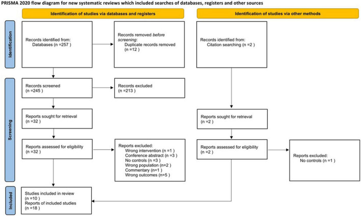
Methods: MEDLINE, Scopus, Cochrane Central Register of Controlled Trials, Web of Science and Embase were systematically searched in September 2020 for case-control studies that reported structural brain MRI in individuals with non-syndromic oral clefts and healthy controls. Studies of syndromic oral clefts were excluded. Two review authors independently screened studies for eligibility, extracted data and assessed risk of bias with the Newcastle-Ottawa Scale. Random effects meta-analyses of mean differences (MDs) and their 95% confidence intervals (95% CI) were performed in order to compare global and regional brain MRI volumes.
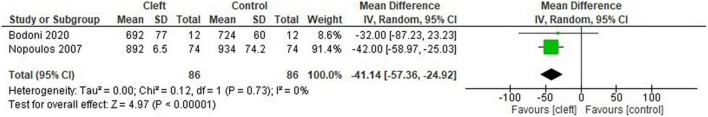
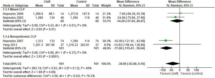
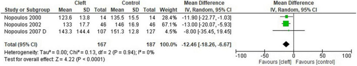
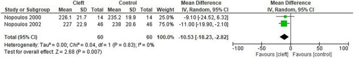
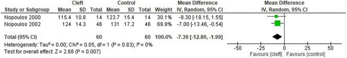
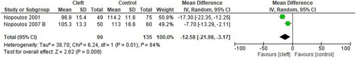
Results: Ten studies from 18 records were included in the review. A total of 741 participants were analyzed. A moderate to high risk of bias was determined for the included studies. The cerebellum (MD: -12.46 cm3, 95% CI: -18.26, -6.67, n = 3 studies, 354 participants), occipital lobes (MD: -7.39, 95% CI: -12.80, -1.99, n = 2 studies, 120 participants), temporal lobes (MD: -10.53 cm3, 95% CI: -18.23, -2.82, n = 2 studies, 120 participants) and total gray matter (MD: -41.14 cm3; 95% CI: -57.36 to -24.92, n = 2 studies, 172 participants) were significantly smaller in the cleft group compared to controls.
Discussion: There may be structural brain differences between individuals with non-syndromic oral clefts and controls based on the available evidence. Improvement in study design, size, methodology and participant selection could allow a more thorough analysis and decrease study heterogeneity.
Keywords: brain; cleft lip; cleft palate; neurodevelopment; neuroimaging.
Copyright © 2022 Sándor-Bajusz, Sadi, Varga, Csábi, Antonoglou and Lohner.
Conflict of interest statement
The authors declare that the research was conducted in the absence of any commercial or financial relationships that could be construed as a potential conflict of interest.
Figures

References
-
- Bjørnland T., Nørholt S. E., Rasmusson L., Sándor G. K. (2021). Nordic Textbook of Oral and Maxillofacial Surgery Munksgaard. Available online at: https://books.google.hu/books?id=aR1rzgEACAAJ (accessed January 26, 2022).
Publication types
LinkOut - more resources
Full Text Sources
Research Materials
Miscellaneous