. 2022 Apr 20;13(22):6496-6501.
doi: 10.1039/d1sc04268h. eCollection 2022 Jun 7.

Quantifying DNA damage on paper sensors via controlled template-independent DNA polymerization

Affiliations

Quantifying DNA damage on paper sensors via controlled template-independent DNA polymerization

Wei Xue et al. Chem Sci. .

Abstract

We report on a paper-based sensor capable of performing template-independent DNA synthesis by terminal deoxynucleotidyl transferase (TdT). Importantly, we observed that TdT efficiently incorporates fluorescently labeled dUTP on to 3'-OH ends of DNA strands in a strictly controllable manner on cellulose paper, in comparison to its distributive mode of DNA synthesis in solution. Due to the high roughness and porous nature of cellulose paper, we attribute this controllable DNA polymerization to the pore confinement effect on the catalytic behaviour of TdT. Taking advantage of this finding, we proposed a paper-assisted TdT (PAT) assay for absolute quantification of alkylated DNA lesions (N7-methylguanine), DNA deamination (cytosine-to-uracil) and DNA oxidation (8-oxo-7,8-dihydroguanine) by combining various DNA glycosylases. This PAT assay provides a low-cost, high throughput and easy to use method for quantifying the absolute levels of various types of DNA lesions, thus making it well-suited for drug development, genotoxicity testing, and environmental toxicology.


Conflict of interest statement

There are no conflicts to declare.

Figures

Fig. 1
Fig. 1. (a) TdT elongation of pA20 initiator. Reactions were carried out with 20 μM FdU, 0.5 μM pA20 and 35 nM TdT at room temperature for various reaction times. Aliquots of the reactions taken at 5, 10, 20, and 30 min were analyzed on a 20% dPAGE (8 M urea) gel. (b) Size distribution of the major products at 5 min. The inset shows that each incorporated FdU generates a new available FdU 3′-OH end for the subsequent incorporation by TdT. SA: streptavidin.
Fig. 2
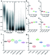
Fig. 2. (a) dPAGE analysis of the products synthesized by TdT at various concentrations of pA20. 35 nM TdT was incubated with 20 μM FdU and pA20 as indicated. Aliquots of the reaction were taken at 30 min. Effect of (b) F-pA20 and (c) P-pA20 concentration on the average DP. Effect of the size of (d) P-pA and (e) F-pA on the average DP. Effect of the sequence of the DNA initiator on the average DP for TdT polymerization (f) on cellulose paper and (g) in solution.
Fig. 3
Fig. 3. (a) Schematic representation of paper-based TdT polymerization in the confined environment provided by the pores of cellulose paper. (b) Average DP plotted against pore size (Φ) of different cellulose papers. (c) TdT elongation of S-pA20 on the surface of nitrocellulose membrane (Millipore HF120). (d) dPAGE analysis of the elongated S-pA20. Reactions were carried out with 20 μM FdU, 50 nM S-pA20 and 35 nM TdT at room temperature for various times. (e) Effect of S-pA20 concentration on the average DP.
Fig. 4
Fig. 4. (a) Strategy of FdU labeling of 7meG lesions by AAG/APE-mediated PAT assay. The DMS-induced damaged base, 7meG is first removed by AAG, and APE cleaves the abasic site, generating 3′-OH ends for subsequent FdU labeling by TdT on paper. (b) In situ labeling of 7meG in DMS-treated ZFL cells. Scale bar: 10 μm. (c) S/B values for paper sensors carried out under different reaction conditions. The extracted genomic DNA was mixed with AAG/APE at 37 °C for 60 min, followed by incubation with TdT and FdU on paper at room temperature for 30 min. The others represent various controls. (d) Absolute quantification of 7meG by mass spectrometric method and PAT assay. ZFL cells (1 × 10⁷) were first treated with varying concentrations of DMS for 30 min before lysis. The error bars in (c) and (d) represent standard deviations of three independent experiments.
Fig. 5
Fig. 5. Working principle of (a) UDG/APE- and (b) FPG/CIAP-mediated PAT assay to measure cytosine damages and 8-oxoG sites on genomic DNA, respectively.
Fig. 6
Fig. 6. (a) DNA repair kinetics. Non-treated (NT) cells were not exposed to DMS. ZFL cells (1× 10⁷) are treated with 2 mM DMS for 30 min at 0 °C, and allowed to repair at DMEM with 10% FBS at 37 °C for 0, 30, and 60 min before lysis. Error bars represent the standard deviation of the replicates. (b) In situ labeling of 7meG in ZFL cells using the AAG/APE-mediated TUNEL assay. DMS-treated cells were cultured at DMEM with 10% FBS at 37 °C for 60 min before labeling. Scale bar: 10 μm.

Similar articles

References

    1. Martinez A. W. Phillips S. T. Whitesides G. M. Anal. Chem. 2010;82:3. doi: 10.1021/ac9013989. - DOI - PubMed
    2. Parolo C. Merkoçi A. Chem. Soc. Rev. 2013;42:450. doi: 10.1039/C2CS35255A. - DOI - PubMed
    3. Gong M. M. Sinton D. Chem. Rev. 2017;117:8447. doi: 10.1021/acs.chemrev.7b00024. - DOI - PubMed
    4. Yang Y. Y. Noviana E. Nguyen M. P. Geiss B. J. Dandy D. S. Henry C. S. Anal. Chem. 2017;89:71. doi: 10.1021/acs.analchem.6b04581. - DOI - PubMed
    5. Zhang Y. Zhang Q. Cheng F. Chang Y. Liu M. Li Y. Chem. Sci. 2021;12:8282. doi: 10.1039/D1SC00795E. - DOI - PMC - PubMed
    1. Liu M. Hui C. Y. Zhang Q. Gu J. Kannan B. Jahanshahi-Anbuhi S. Filipe C. D. M. Brennan J. D. Li Y. Angew. Chem., Int. Ed. 2016;55:2709. doi: 10.1002/anie.201509389. - DOI - PubMed
    2. Bi S. Ji B. Zhang Z. Zhu J. Chem. Sci. 2013;4:1858. doi: 10.1039/C3SC00043E. - DOI
    1. Xu G. Nolder D. Reboud J. Oguike M. C. van Schalkwyk D. A. Sutherland C. J. Cooper J. M. Angew. Chem., Int. Ed. 2016;55:15250. doi: 10.1002/anie.201606060. - DOI - PMC - PubMed
    2. Tang Y. Lu B. Zhu Z. Li B. Chem. Sci. 2018;9:760. doi: 10.1039/C7SC03190D. - DOI - PMC - PubMed
    1. Ahn H. Batule B. S. Seok Y. Kim M. G. Anal. Chem. 2018;90:10211. doi: 10.1021/acs.analchem.8b01309. - DOI - PubMed
    2. Rohrman B. Richards-Kortum R. Lab Chip. 2012;12:3082. doi: 10.1039/C2LC40423K. - DOI - PMC - PubMed
    1. Linnes J. Rodriguez N. Liu L. Klapperich C. Biomed. Microdevices. 2016:18. - PMC - PubMed
    2. Tang R. Yang H. Gong Y. You M. Liu Z. Choi J. R. Wen T. Qu Z. Mei Q. Xu F. Lab Chip. 2017;17:1270. doi: 10.1039/C6LC01586G. - DOI - PubMed