. 2021:2:86-100.
doi: 10.1109/ojnano.2021.3119913. Epub 2021 Oct 14.

Design Approaches and Computational Tools for DNA Nanostructures

Affiliations

Design Approaches and Computational Tools for DNA Nanostructures

Heeyuen Koh et al. IEEE Open J Nanotechnol. 2021.

Abstract

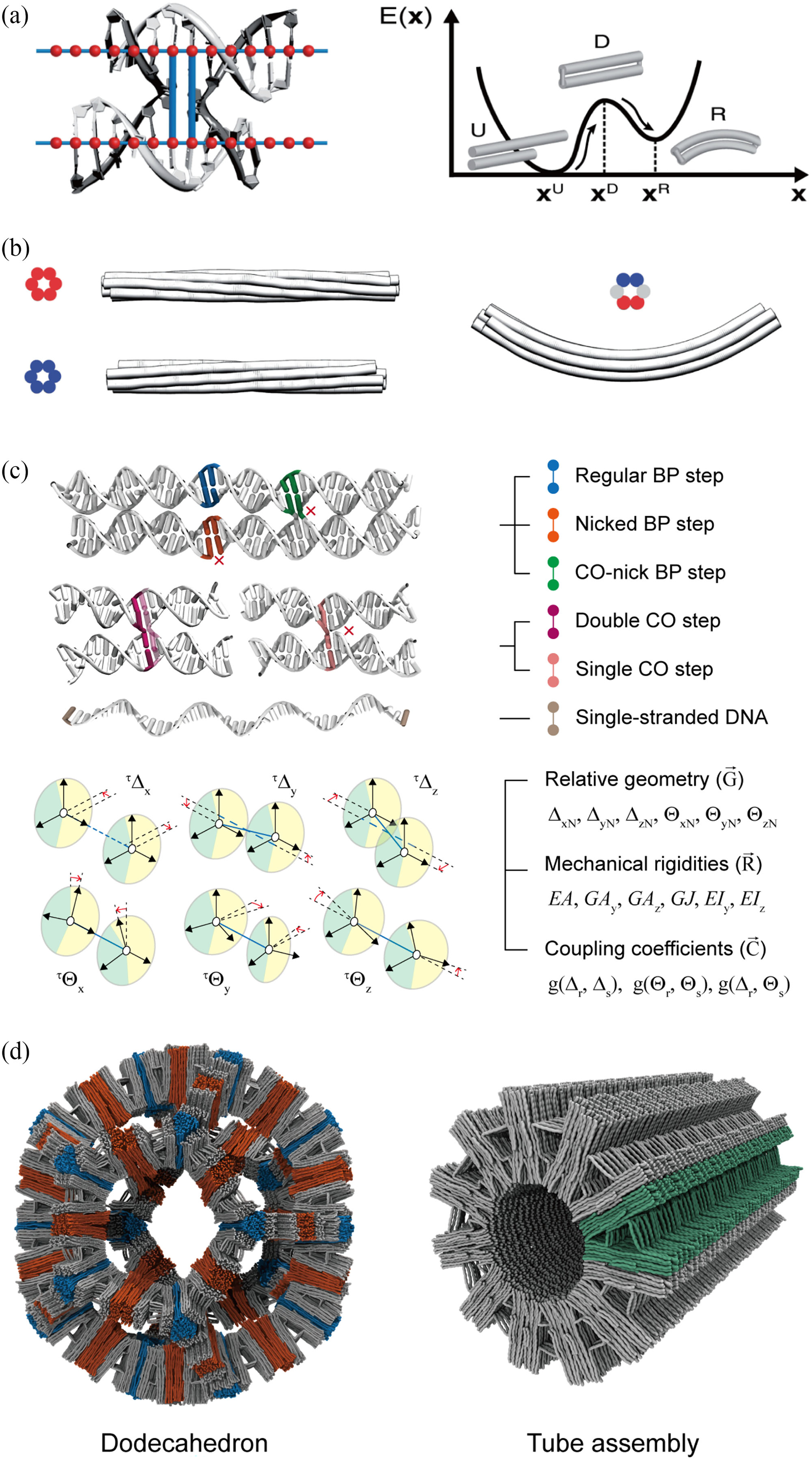
Designing a structure in nanoscale with desired shape and properties has been enabled by structural DNA nanotechnology. Design strategies in this research field have evolved to interpret various aspects of increasingly more complex nanoscale assembly and to realize molecular-level functionality by exploring static to dynamic characteristics of the target structure. Computational tools have naturally been of significant interest as they are essential to achieve a fine control over both shape and physicochemical properties of the structure. Here, we review the basic design principles of structural DNA nanotechnology together with its computational analysis and design tools.

Keywords: DNA; computational tools; design principle; nanotechnology.


Figures

FIGURE 1.
FIGURE 1.
Example of DNA origami structure: (A) General process to complete origami from design stage to realize the structure in solution, (B) Examples of motifs in DNA nanostructure, (C) Examples of modules. Left, displacement reaction of DNA tile designed as modules [83]. Right, examples of motif design [59]. Crystal cell aligned with the crystallographic structure PDB ID: 3GBI (light blue) and overall crystal structure of PDB ID: 3GBI, respectively. Scale bars are 1 nm (left) and 10 nm (right). Both figures are distributed in terms of the Creative Commons CC BY license.
FIGURE 2.
FIGURE 2.
Development of DNA origami computational tools. (A) Blunt end using 3-helix motif design using GIDEON. Reproduced from [105] with permission from The Royal Society of Chemistry. (B) SARSE [94]. Reprinted with permission. Copyright 2008, American Chemical Society. (C) Screenshot from TIAMAT [95]. (D) Screenshot from caDNAno [96]. (E) Stanford bunny made with v-Helix [87]. (F) Wire-framed polyhedron by DAEDALUS [102]. (G) Vertex connectivity designed by TALOS[103]. (H) 2D wire-framed structure by PERDIX [104]. (I) Banner of the webservice for tacoxDNA [106] from http://tacoxdna.sissa.it/. (J) ATHENA [107].
FIGURE 3.
FIGURE 3.
Atomistic molecular dynamics simulation. (A) Holliday junction in atomic simulation [38]. Copyright 2016 National Academy of Science. (B) Conformation of honeycomb structure [38]. Copyright 2016 National Academy of Science. (C) Schematic representation of a six-helix bundle. Reprinted from [8] with the permission. Copyright 2019 American Chemical Society. (D) Snapshot showing an equilibrated configuration of the six-helix bundle with gaps. Red-colored regions indicate the gap positions. Reprinted from [8] with the permission. Copyright 2019 American Chemical Society.
FIGURE 4.
FIGURE 4.
Coarse-grained (CG) molecular dynamics simulation. (A) Configuration change from oxDNA to oxDNA2 for major-minor groove (top). Reprinted from [122] with the permission of AIP Publishing. Specific potential energy modeling for dsDNA (bottom). Reproduced from [99] with the permission from the PCCP Owner Societies. (B) DNA walker simulated by oxDNA [40]. Copyright 2017, Oxford University Press. (C) Linkage system in oxDNA simulation. Reproduced from [37] with the permission from the Royal Society of Chemistry. (D) Schematic representation of CG particles [116]. Copyright 2014, American Chemical Society. (E) Multiscale prediction using MrDNA [97]. Copyright 2020, Oxford University Press.
FIGURE 5.
FIGURE 5.
Finite-element-based structural analysis. (A) DNA double helices as isotropic elastic rods with rigid crossovers. Reproduced with permission, Copyright 2011, Oxford University Press [42]. (B) Example deformations induced by the mismatch between neighboring DNA helices by (red) insertions and (blue) deletions for a honeycomb lattice bundle [42]. Copyright 2011, Oxford University Press. (C) Classification and characterization of structural motifs in DNA nanostructures. Reproduced with permission, Copyright 2011, Oxford University Press [43], Copyright 2021, American Chemical Society [131]. (D) Prediction results of giga-dalton scale hierarchical assemblies of dodecahedron and tube, Copyright 2021, American Chemical Society [131].
FIGURE 6.
FIGURE 6.
Theoretical models. (A) Orthogonal set of vectors defined at each point of a rod (grey-purple). s is the arc-length of the rod, and the subset with red dashed line indicates the unit vector definition based on the groove condition so that the unit vector set has twist angle along the double helix [135]. Different conditions of unit vector set along the rod causes the perturbation on the strain energy to consider the unique strain energy condition of double helix. Reprinted from [133] with the permission. Copyright 2019, by the American Physical Society. (B) Schematic cartoons for topological changes due to the difference in entropic cost. Reprinted from [125] with the permission of AIP Publishing.

Similar articles

Cited by

References

    1. Seeman NC, “Nucleic acid junctions and lattices,” J. Theor. Biol, vol. 99, no. 2, pp. 237–247, 1982. - PubMed
    1. Rothemund PWK, “Folding DNA to create nanoscale shapes and patterns,” Nature, vol. 440, no. 7082, pp. 297–302, 2006. - PubMed
    1. Pfeifer W and Saccà B,“From nano to macro through hierarchical self-assembly: The DNA paradigm,” ChemBioChem, vol. 17, pp. 1063–1080, 2016. - PubMed
    1. Mathieu F, Liao S, Kopatsch J, Wang T, Mao C, and Seeman NC, “Six-helix bundles designed from DNA,” Nano Lett, vol. 5, no. 4, pp. 661–665, 2005. - PMC - PubMed
    1. Dietz H, Douglas SM, and Shih WM, “Folding DNA into twisted and curved nanoscale shapes,” Sci. (80-.), vol. 325, no. 5941, pp. 725–730, 2009. - PMC - PubMed

LinkOut - more resources