

Malignant Transformation of Recurrent Residual Cerebellopontine Angle Epidermoid Tumor: Significance of Clinical Vigilance and Long-Term Surveillance
- PMID: 35756906
- PMCID: PMC9232296
- DOI: 10.1055/a-1858-7483
Malignant Transformation of Recurrent Residual Cerebellopontine Angle Epidermoid Tumor: Significance of Clinical Vigilance and Long-Term Surveillance
Abstract
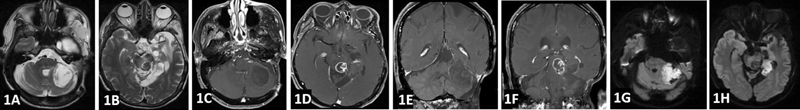
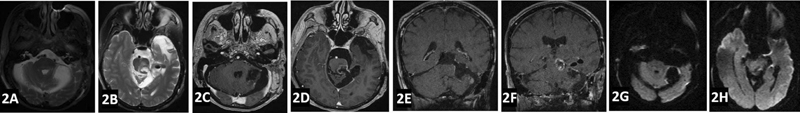
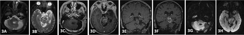
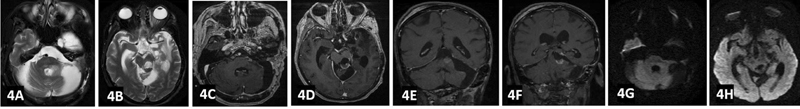
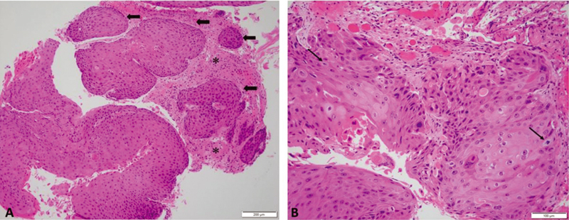
Epidermoid tumors (ET) are slow-growing masses where malignant transformations occur extremely rarely. Malignant transformation warning signs are the rapid-onset, progression, and recurrence of symptoms. The radiologic evidence for malignant transformation is contrast enhancement with rapid growth, observed with magnetic resonance imaging (MRI) or computed tomography scans. Here, we provide a case report of a 68-year-old woman with a long-standing history of left-sided cerebellopontine angle ET who presented with a recent worsening of symptoms, and MRI observation of new ET contrast enhancement. Surgical re-exploration and histopathologic confirmation are mandatory in this setting of recent symptom worsening and MRI observation of rapid mass growth.
Keywords: brain tumor; cerebellopontine angle; epidermoid tumor; malignant transformation; rapid progression; recurrence; squamous cell carcinoma.
The Author(s). This is an open access article published by Thieme under the terms of the Creative Commons Attribution-NonDerivative-NonCommercial License, permitting copying and reproduction so long as the original work is given appropriate credit. Contents may not be used for commercial purposes, or adapted, remixed, transformed or built upon. ( https://creativecommons.org/licenses/by-nc-nd/4.0/ ).
Conflict of interest statement
Conflict of Interest None declared.
Figures
Similar articles
-
[Malignant Transformation of Cerebellopontine Angle Epidermoid Cyst:A Case Report].No Shinkei Geka. 2019 Nov;47(11):1173-1178. doi: 10.11477/mf.1436204095. No Shinkei Geka. 2019. PMID: 31761779 Japanese.
-
Malignant squamous degeneration of a cerebellopontine angle epidermoid tumor. Case report.J Neurosurg. 2002 Nov;97(5):1237-43. doi: 10.3171/jns.2002.97.5.1237. J Neurosurg. 2002. PMID: 12450053 Review.
-
Radio-pathological characteristics of malignant transformation of an epidermoid cyst in the cerebellopontine angle: A case report.Surg Neurol Int. 2022 Apr 8;13:135. doi: 10.25259/SNI_1226_2021. eCollection 2022. Surg Neurol Int. 2022. PMID: 35509542 Free PMC article.
-
Malignant transformation of an epidermoid cyst in the cerebellopontine angle.J Korean Neurosurg Soc. 2012 Aug;52(2):148-51. doi: 10.3340/jkns.2012.52.2.148. Epub 2012 Aug 31. J Korean Neurosurg Soc. 2012. PMID: 23091675 Free PMC article.
-
Cerebellar Squamous Cell Carcinoma Due to Malignant Transformation of Cerebellopontine Angle Epidermoid Cyst, Report an Interesting Case and Review the Literature.Prague Med Rep. 2019;120(2-3):95-102. doi: 10.14712/23362936.2019.14. Prague Med Rep. 2019. PMID: 31586508 Review.
References
-
- Link M J, Cohen P L, Breneman J C, Tew J M., Jr Malignant squamous degeneration of a cerebellopontine angle epidermoid tumor. Case report. J Neurosurg. 2002;97(05):1237–1243. - PubMed
-
- Nakao Y, Nonaka S, Yamamoto T. Malignant transformation 20 years after partial removal of intracranial epidermoid cyst–case report. Neurol Med Chir (Tokyo) 2010;50(03):236–239. - PubMed
-
- Hamlat A, Hua Z F, Saikali S. Malignant transformation of intra-cranial epithelial cysts: systematic article review. J Neurooncol. 2005;74(02):187–194. - PubMed
-
- Tamura K, Aoyagi M, Wakimoto H. Malignant transformation eight years after removal of a benign epidermoid cyst: a case report. J Neurooncol. 2006;79(01):67–72. - PubMed
Publication types
LinkOut - more resources
Full Text Sources
