. 2022 Jun 8:10:887393.
doi: 10.3389/fcell.2022.887393. eCollection 2022.

Trunk Neural Crest Migratory Position and Asymmetric Division Predict Terminal Differentiation

Affiliations

Trunk Neural Crest Migratory Position and Asymmetric Division Predict Terminal Differentiation

Zain Alhashem et al. Front Cell Dev Biol. .

Abstract

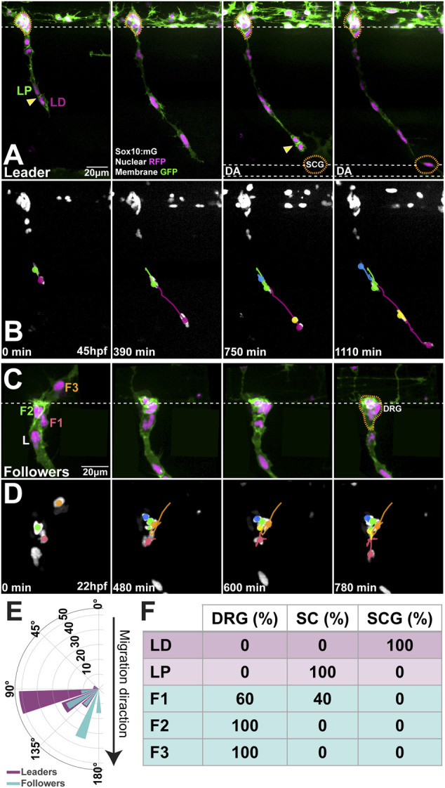
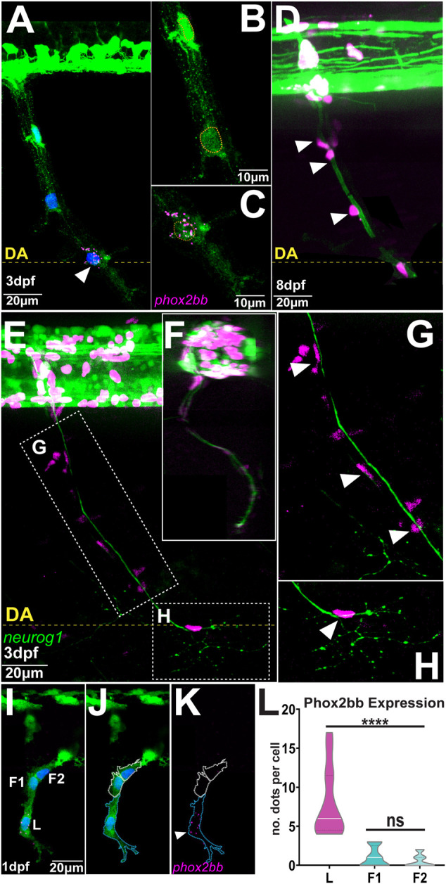
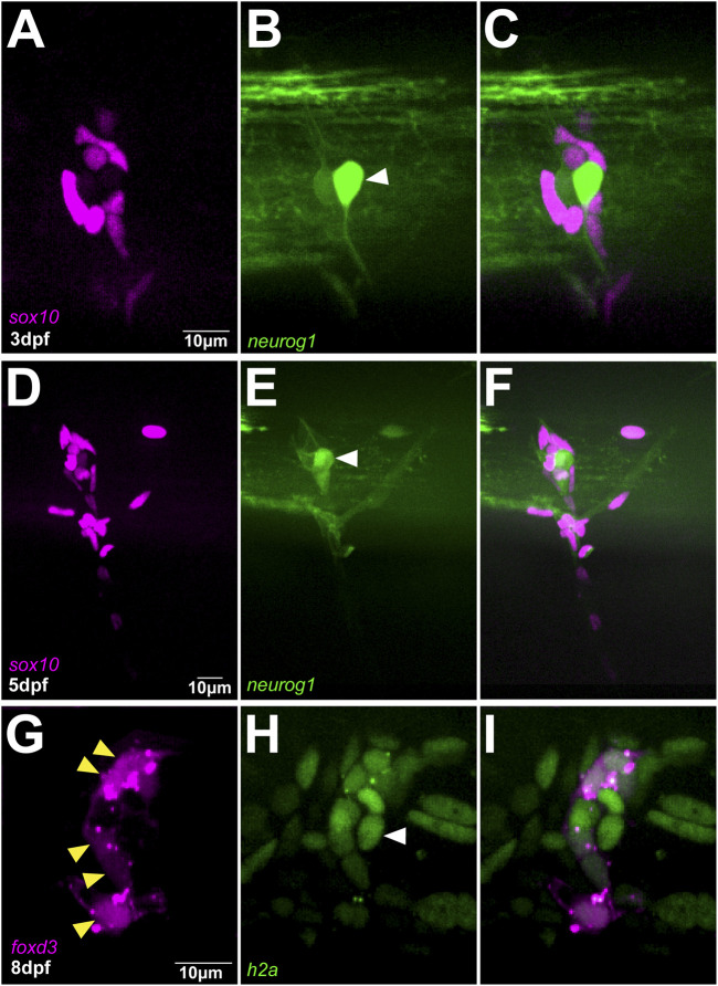
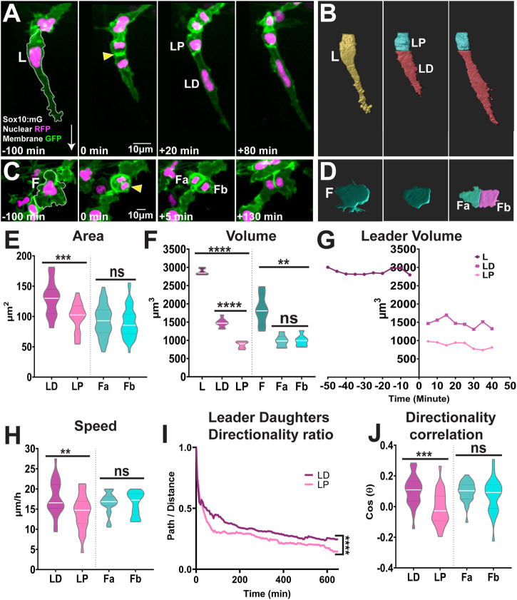
The generation of complex structures during embryogenesis requires the controlled migration and differentiation of cells from distant origins. How these processes are coordinated and impact each other to form functional structures is not fully understood. Neural crest cells migrate extensively giving rise to many cell types. In the trunk, neural crest cells migrate collectively forming chains comprised of cells with distinct migratory identities: one leader cell at the front of the group directs migration, while followers track the leader forming the body of the chain. Herein we analysed the relationship between trunk neural crest migratory identity and terminal differentiation. We found that trunk neural crest migration and fate allocation is coherent. Leader cells that initiate movement give rise to the most distal derivativities. Interestingly, the asymmetric division of leaders separates migratory identity and fate. The distal daughter cell retains the leader identity and clonally forms the Sympathetic Ganglia. The proximal sibling migrates as a follower and gives rise to Schwann cells. The sympathetic neuron transcription factor phox2bb is strongly expressed by leaders from early stages of migration, suggesting that specification and migration occur concomitantly and in coordination. Followers divide symmetrically and their fate correlates with their position in the chain.

Keywords: asymmetric division; collective migration; dorsal root ganglia; fate; leader; neural crest; schwann cell; sympathetic ganglia.


Conflict of interest statement

The authors declare that the research was conducted in the absence of any commercial or financial relationships that could be construed as a potential conflict of interest.

Figures

FIGURE 1
FIGURE 1
Migratory position predicts differentiation outcome (A,B) Selected frames of (A) live imaging and (B) cell tracks starting at 45hpf of a Sox10:mG embryo at the level of somite 20 (C,D) Selected frames of (C) live imaging and (D) cell tracks of Sox10:mG between 22 and 35hpf at the level of somite 9. L: leader, F1: first follower, F2: second follower, F3: third follower (E) Orientation of the division plane (leaders n = 27, followers n = 43) (F) Derivatives formed by each cell in the migratory chain (n = 14 chains). Anterior to the left, dorsal top. Yellow arrows indicate divisions; orange dotted line DRG and SCG positions. LD leader’s distal and LP leader’s proximal daughters. DRG: dorsal root ganglia; SCG: sympathetic chain ganglia; SC: Schwann cell. Time in minutes after the beginning of the movie.
FIGURE 2
FIGURE 2
Phox2bb is predominantly expressed in leader cells (A–C) phox2bb expression at 3dpf (B) Enlargements of follower and (C) leader cells. Sox10 in green, phox2bb in magenta and nuclei in blue in panel A and highlighted with orange dotted lines in B-C (D) Confocal image of Sox10:mG embryo at 8dpf. White arrowheads indicate TNC cells showing Schwann cell morphology (E–H) Confocal images of Sox10:mG; neurogenin1:GFP at 3dpf showing sensory neuron axon extension from DRG to SCG lateral view in (E) and transversal section in (F) (G) Enlargement showing TNC followers localising around the axon in position of Schwann cells indicated by white arrowheads and (H) Enlargement showing single leader cell in the position of SCG (I–K) Confocal image of Sox10:GFP embryo at 1dpf showing expression of phox2bb in leader cell. L: Leader, F1 and F2: first and second followers. White arrowheads indicate leader. Blue and white dotted lines mark the outlines of leader and follower cells respectively (L) Quantification of phox2bb expression (n = 14 chains; Welch’s t test, L vs. F1 and F2 p < 0.0001, F1 vs. F2 p = 0.2566). Anterior to the left, dorsal top. DA: Dorsal Aorta.
FIGURE 3
FIGURE 3
DRG is exclusively formed by followers (A–C) Confocal images of Sox10:mG; neurogenin1:GFP at 3dpf showing the differentiation of a follower cell into the primary neuron of DRG indicated by white arrowhead (D–F) Confocal images of in Sox10:mG; neurogenin1:GFP embryo at 5dpf showing a more mature DRG (G–I) Confocal images of FoxD3:mCherry; H2aFVA:H2A-GFP embryo at 8dpf showing the peripheral followers expressing FoxD3, yellow arrowheads, and the primary neuron marked by the absence of FoxD3 expression, white arrowhead.
FIGURE 4
FIGURE 4
Leaders TNC divide asymmetrically while followers divide symmetrically (A,B) Confocal images of Sox10:mG (A) and 3D rendering (B) of leader asymmetric division into distal and proximal daughters. L: Leader, LD: Leader Distal, LP: Leader Proximal; white arrow indicates direction of migration and yellow arrowhead points to the dividing cell (C,D) Confocal images of Sox10:mG (C) and 3D rendering (D) of follower symmetric division. F: Follower, Fa and Fb Follower’s daughters (E) Quantification of cell area of leaders’ and followers’ daughters quantified immediately after division (n = 25 for leaders and n = 44 for followers; Unpaired t test, p = 0.0001 for LD vs. LP and p = 0.5852 for Fa vs. Fb) (F) Quantification of cell volume of leaders, followers and their daughters after division (n = 10 divisions for leaders and n = 10 divisions for followers; Brown-Forsythe and Welch ANOVA tests, p < 0.0001 for L vs. LD and LP, p < 0.0001 for LD vs. LP, p = 0.0012 for F vs. Fa and Fb, p > 0.9999 for Fa vs. Fb) (G) Cell volume over time of a representative leader before and after division (H) Quantification of speed of movement of leaders’ and followers’ daughters (n = 25 for leaders and n = 44 for followers; Unpaired t test, p = 0.0072 for LD vs. LP, p = 0.8690 for Fa vs. Fb) (I) Directionality ratio of leaders’ daughters (n = 25 divisions; Simple linear regression, p < 0.0001 for LD vs. LP) (J) Directionality correlation of leaders’ and followers’ daughters (n = 25 for leaders and n = 44 for followers; Welch’s t test, p = 0.0004 for LD vs. LP, p = 0.2798 for Fa vs. Fb).

References

    1. Adameyko I., Lallemend F., Aquino J. B., Pereira J. A., Topilko P., Müller T., et al. (2009). Schwann Cell Precursors from Nerve Innervation Are a Cellular Origin of Melanocytes in Skin. Cell 139, 366–379. 10.1016/j.cell.2009.07.049 - DOI - PubMed
    1. Alhashem Z., Feldner-Busztin D., Revell C., Alvarez-Garcillan Portillo M., Camargo-Sosa K., Richardson J., et al. (2022). Notch Controls the Cell Cycle to Define Leader versus Follower Identities during Collective Cell Migration. eLife 11, e73550. 10.7554/eLife.73550 - DOI - PMC - PubMed
    1. Alhashem Z., Portillo M. A.-G., Htun M. R., Gauert A., Montecinos L. B., Härtel S., et al. (2021). “Zebrafish Neural Crest: Lessons and Tools to Study In Vivo Cell Migration,” in The Epithelial-To Mesenchymal Transition: Methods and Protocols. Editors Campbell K., Theveneau E. (New York, NY: Springer US; ), 79–106. Methods in Molecular Biology. 10.1007/978-1-0716-0779-4_9 - DOI - PubMed
    1. An M., Luo R., Henion P. D. (2002). Differentiation and Maturation of Zebrafish Dorsal Root and Sympathetic Ganglion Neurons. J. Comp. Neurol. 446, 267–275. 10.1002/cne.10214 - DOI - PubMed
    1. Baggiolini A., Varum S., Mateos J. M., Bettosini D., John N., Bonalli M., et al. (2015). Premigratory and Migratory Neural Crest Cells Are Multipotent In Vivo . Cell Stem Cell 16, 314–322. 10.1016/j.stem.2015.02.017 - DOI - PubMed

LinkOut - more resources