. 2022 Jun 8:10:879057.
doi: 10.3389/fcell.2022.879057. eCollection 2022.

Inhibition of the Protein Arginine Methyltransferase PRMT5 in High-Risk Multiple Myeloma as a Novel Treatment Approach

Affiliations

Inhibition of the Protein Arginine Methyltransferase PRMT5 in High-Risk Multiple Myeloma as a Novel Treatment Approach

Philip Vlummens et al. Front Cell Dev Biol. .

Abstract

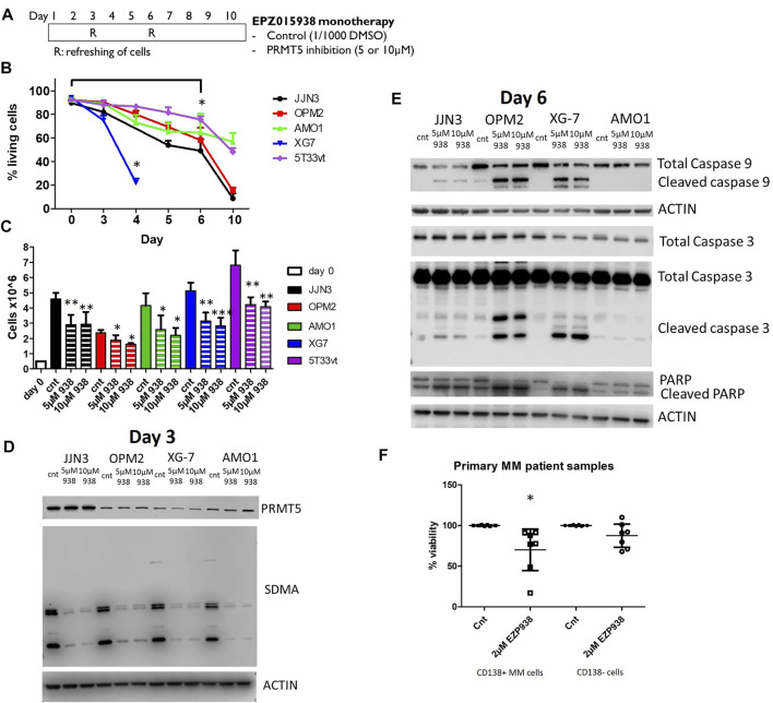
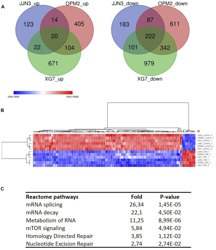
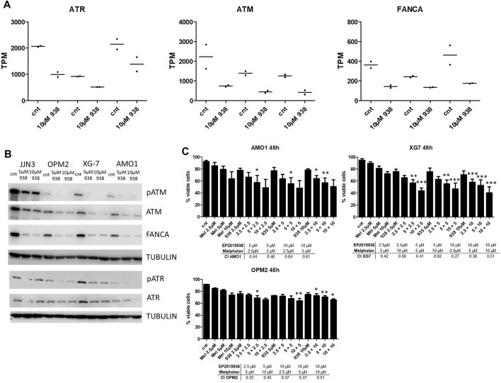
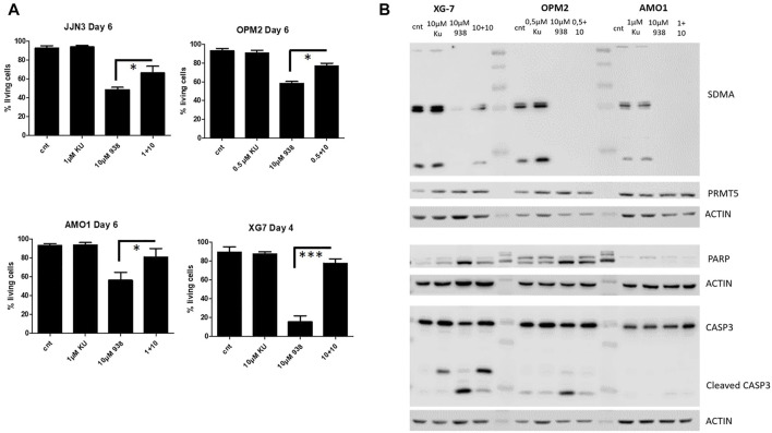
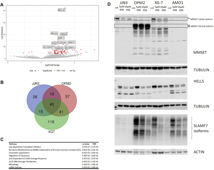
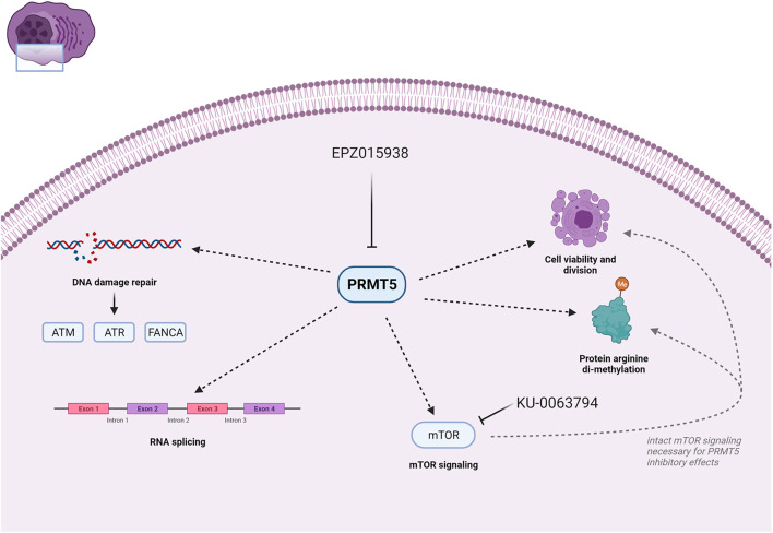
Multiple myeloma (MM) is an incurable clonal plasma cell malignancy. Subsets of patients have high-risk features linked with dismal outcome. Therefore, the need for effective therapeutic options remains high. Here, we used bio-informatic tools to identify novel targets involved in DNA repair and epigenetics and which are associated with high-risk myeloma. The prognostic significance of the target genes was analyzed using publicly available gene expression data of MM patients (TT2/3 and HM cohorts). Hence, protein arginine methyltransferase 5 (PRMT5) was identified as a promising target. Druggability was assessed in OPM2, JJN3, AMO1 and XG7 human myeloma cell lines using the PRMT5-inhibitor EPZ015938. EPZ015938 strongly reduced the total symmetric-dimethyl arginine levels in all cell lines and lead to decreased cellular growth, supported by cell line dependent changes in cell cycle distribution. At later time points, apoptosis occurred, as evidenced by increased AnnexinV-positivity and cleavage of PARP and caspases. Transcriptome analysis revealed a role for PRMT5 in regulating alternative splicing, nonsense-mediated decay, DNA repair and PI3K/mTOR-signaling, irrespective of the cell line type. PRMT5 inhibition reduced the expression of upstream DNA repair kinases ATM and ATR, which may in part explain our observation that EPZ015938 and the DNA-alkylating agent, melphalan, have combinatory effects. Of interest, using a low-dose of mTOR-inhibitor, we observed that cell viability was partially rescued from the effects of EPZ015938, indicating a role for mTOR-related pathways in the anti-myeloma activity of EPZ015938. Moreover, PRMT5 was shown to be involved in splicing regulation of MMSET and SLAMF7, known genes of importance in MM disease. As such, we broaden the understanding of the exact role of PRMT5 in MM disease and further underline its use as a possible therapeutic target.

Keywords: DNA repair; PRMT5; RNA splicing; epigenetics; myeloma.


Conflict of interest statement

The authors declare that the research was conducted in the absence of any commercial or financial relationships that could be construed as a potential conflict of interest.

Figures

FIGURE 1
FIGURE 1
(A): Schematic representation of EPZ01938 therapy in HMCLs. Cells were incubated with either compound or DMSO as placebo. Cells were refreshed at predetermined intervals. At timepoints R cells were gently harvested, counted and replated at the initial cellular concentration (0.1 × 10^6 cells/ml) on day 3 and 6 by in fresh medium to which either DMSO or EPZ015938 was added. (B): Viability assay of HMCLs upon treatment with EPZ015938. Viability was assessed by trypane blue staining at depicted timepoints for the selected HMCLs. Treated samples were compared against control samples. Error bars depict mean values ± SD; * denotes p < 0.05 (n > 3) (C): Cumulative cell counts of living cells for each HMCL when cultured with or without 5 and 10 µM EPZ015938 after 3 days of treatment. Treated samples were compared against control samples. Error bars depict mean values ± SD; */**/*** denotes p < 0.05, p < 0.01 and p < 0.001 respectively (n > 3) (D): Western blot staining for PRMT5 and SDMA protein levels was performed after 3 days of EPZ015938 treatment on JJN3, OPM2, XG7 and AMO1 cells. Actin was used as a loading control. One experiment representative of 3 experiments performed is shown (n = 3). (E): Western blot of pro-apoptotic proteins caspase 9, 3 and PARP after 3 days of treatment with EOZ015938. Actin was added as loading control. One representative experiment is shown (n = 3). (F): Effect of PRMT5 inhibitor treatment on primary human CD138 + MM cells and CD138- microenvironment. Mononuclear cells from 7 MM patients were treated with the indicated concentration for 4 days, and the percentage of viable CD138 + plasma cells and CD138- cells were determined by flow cytometry. Results are expressed as the relative viability compared with control. * denotes p < 0.05 compared to control cells.
FIGURE 2
FIGURE 2
(A): Deregulated genes after PRMT5 inhibition were identified using RNA-seq and analysed for JJN3, OPM2 and XG7 cells separately due to possible cell type specific events. Venn diagram analysis shows the presence of a specific set of common up- (left) or downregulated (right) genes per cell line type (n = 2 × 2 per cell type/treatment). (B): Unsupervised clustering of analysed samples used for RNA-seq based on common deregulated genes (n = 242), clearly segregating according to treatment type. (C): Overview of reactome pathway analysis output (selected pathways are shown) with fold changes and p-values per pathway, after multiple testing correction.
FIGURE 3
FIGURE 3
(A): TPM values for ATR, ATM and FANCA for both control and EPZ015938 cells, showing decrease in transcript abundancy following PRMT5 inhibition in all 3 cell lines studied (n = 2 for each cell line and condition, no error bars are shown). (B): Western blot analysis was performed on JJN3, OPM2, XG7 and AMO1 cell lysates to evaluate ATM/ATR and FANCA protein levels upon PRMT5 inhibition for 3 consecutive days. Tubulin was used as a loading control. One experiment representative of 3 experiments performed is shown (n = 3). (C): Viability assay using Annexin/7AAD-staining of HMCLs after treatment with EPZ015938, melphalan or combination therapy at depicted concentrations. Samples were analysed using an ANOVA selected pairs test. to evaluate differences between different treatment settings and against control samples and samples treated with only EPZ015938 or melphalan. Error bars represent mean +SD; */**/*** denotes p < 0.05, p < 0.01 and p < 0.001 respectively (n = 3). Synergy was evaluated for between both compounds by calculation of the combination index (CI) for significant combinations, with a CI < 1 showing a synergistic effect.
FIGURE 4
FIGURE 4
(A): Viability assay of HMCls upon treatment of JJN3, OPM2, AMO1 and XG7 cells with EPZ015938, KU-0063794 or combination therapy after predefined timepoints. Cellular viability was assessed using AnnexinV/7AAD staining. Samples were analysed using an T—test with selected pairs test, significance was evaluated between different treatment settings and compared to control. Error bars depict mean ± SD; */**/*** denotes p < 0.05, p < 0.01 and p < 0.001 respectively (n = 3). (B): Western blot analysis of SDMA levels, global PRMT5 levels and pro-apoptotic proteins caspase 9, 3 and PARP in XG7, OPM2 and AMO1 cells. Cells were treated with EPZ015938, KU-0063794 or combination therapy. Control samples were included for comparison. Actin was used as a loading control. One experiment representative of 3 experiments performed is shown (n = 3).
FIGURE 5
FIGURE 5
(A): Intron retention due to PRMT5 inhibition was evaluated by re-analysis of RNA-seq data (as described earlier) using IRFinder (n = 2 for each condition and per cell line). Volcano plot shows IRFinder output in JJN3 cells, identifying different gene targets affected by intron retention. Only the plot for JJN3 is shown here, other plots are shown in the supplementary data. (B): Venn diagram presentation of common genes affected by intron retention (n = 2 × 2 per HMCL/treatment condition). A set of 45 common affected genes were identified. Samples were analysed per cell line type to account for cell type specific effects. (C): Reactome enrichment analysis was subsequently performed using the 45 common genes. Selected pathways that were deregulated are shown. (D): Western blot analysis of identified targets MMSET/WHSC1 HELLS and SLAMF7 in JJN3, OPM2, XG7 and AMO1 cells. For MMSET, * depicts the wild type MMSET isoform where § depicts the aberrant MMSET protein affected by t (4; 14), which is present in OPM2 cells. Tubulin and actin were used as loading control. One experiment representative of 3 experiments performed is shown (n = 3).
FIGURE 6
FIGURE 6
Schematic representation of PRMT5 function and effects upon treatment as evaluated in this study. PRMT5 inhibition by using i.e., EPZ015938 leads to decreased DNA damage repair through a decrease in ATM/ATR and FANCA transcript and protein levels. Moreover, the function of PRMT5 as a regulator of mRNA splicing also plays a role in MM cellular processes and survival. Lastly, our study shows that mTOR signalling is important for PRMT5 function and has effects on cell viability, division and overall arginine di-methylation. Created with BioRender.

Similar articles

Cited by

References

    1. Banasavadi-Siddegowda Y. K., Welker A. M., An M., Yang X., Zhou W., Shi G., et al. (2018). PRMT5 as a Druggable Target for Glioblastoma Therapy. Neuro Oncol. 20 (6), 753–763. 10.1093/neuonc/nox206 - DOI - PMC - PubMed
    1. Barlogie B., Tricot G., Rasmussen E., Anaissie E., van Rhee F., Zangari M., et al. (2006). Total Therapy 2 without Thalidomide in Comparison with Total Therapy 1: Role of Intensified Induction and Posttransplantation Consolidation Therapies. Blood 7(7), 2633–2638. 10.1182/blood-2005-10-4084 - DOI - PubMed
    1. Binder M., Rajkumar S. V., Ketterling R. P., Dispenzieri A., Lacy M. Q., Gertz M. A., et al. (2019). Substratification of Patients with Newly Diagnosed Standard‐risk Multiple Myeloma. Br. J. Haematol. 185 (2), 254–260. 10.1111/bjh.15800 - DOI - PubMed
    1. Broyl A., Hose D., Lokhorst H., de Knegt Y., Peeters J., Jauch A., et al. (2010). Gene Expression Profiling for Molecular Classification of Multiple Myeloma in Newly Diagnosed Patients. Blood 116 (14), 2543–2553. 10.1182/blood-2009-12-261032 - DOI - PubMed
    1. Caprio C., Sacco A., Giustini V., Roccaro A. M. (2020). Epigenetic Aberrations in Multiple Myeloma. Cancers 12 (10), 2996. 10.3390/cancers12102996 - DOI - PMC - PubMed