Review
. 2022 Jun 9:10:876815.
doi: 10.3389/fcell.2022.876815. eCollection 2022.

The Intimate Connection Between Lipids and Hedgehog Signaling

Affiliations
Review

The Intimate Connection Between Lipids and Hedgehog Signaling

Thi D Nguyen et al. Front Cell Dev Biol. .

Abstract

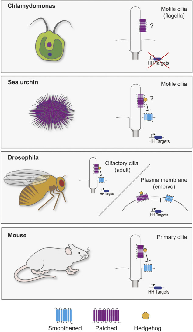
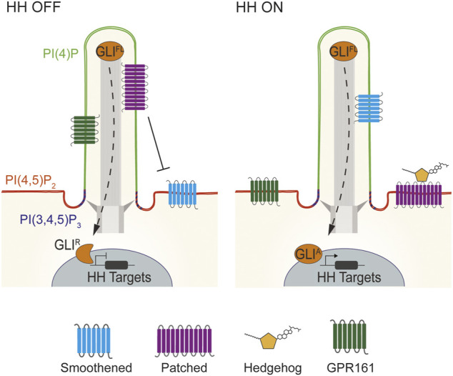
Hedgehog (HH) signaling is an intercellular communication pathway involved in directing the development and homeostasis of metazoans. HH signaling depends on lipids that covalently modify HH proteins and participate in signal transduction downstream. In many animals, the HH pathway requires the primary cilium, an organelle with a specialized protein and lipid composition. Here, we review the intimate connection between HH signaling and lipids. We highlight how lipids in the primary cilium can create a specialized microenvironment to facilitate signaling, and how HH and components of the HH signal transduction pathway use lipids to communicate between cells.

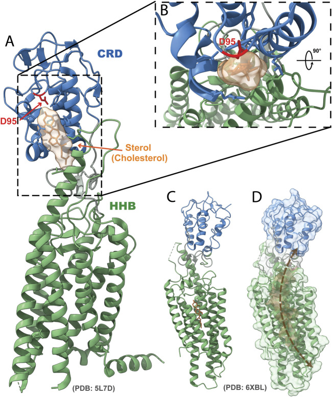
Keywords: cholesterolyation; cilia; development; intercellular signaling; sterols.


Conflict of interest statement

The authors declare that the research was conducted in the absence of any commercial or financial relationships that could be construed as a potential conflict of interest.

Figures

FIGURE 1
FIGURE 1
Cilia and Hedgehog signaling throughout evolution. Though cilia are highly conserved, the reliance of HH signaling on the cilium varies through evolution. The green alga Chlamydomonas reinhardtii genome possesses two homologs of PTCH (Cre02.g093500 and Cre12.g496350), but not other components of the HH pathway. It will be interesting to determine whether either acts at the Chlamydomonas flagella. In sea urchin embryos, SMO localizes to cilia to activate HH signaling for mesoderm specification (Warner et al., 2014). Different tissues in Drosophila transduce HH signals with or without cilia. Most Drosophila cells lack cilia and transduce HH signals via Smo at the plasma membrane (Zhu et al., 2003; Jia et al., 2004). However, olfactory sensory neuron cilia in the adult fly brain signal through ciliary Smo (Kuzhandaivel et al., 2014). Vertebrates require primary cilia to transduce HH signals (Bangs and Anderson, 2017). Defects in ciliary transport or structure cause a wide range of HH-related phenotypes (Reiter and Leroux, 2017).
FIGURE 2
FIGURE 2
Lipid domains within vertebrate cilia. Vertebrate HH signaling depends on the tightly coordinated trafficking of pathway components into and out of cilia. In the absence of HH signals, PTCH1 localizes to cilia and represses SMO. Consequently, GLI transcription factors are proteolytically processed to their repressor state to inhibit HH target genes. HH binding to PTCH1 leads to SMO accumulation in cilia, GPR161 exit, and GLI activator formation to induce HH target gene expression (Bangs and Anderson, 2017; Kong et al., 2019). Specialized membrane domains within mouse and human cilia allow for HH signal transduction. Mouse and human cilia are enriched in the phosphoinositide PI(4)P, whereas PI(4,5)P2 is enriched outside of the cilium. Defects in the distribution of these two lipids causes mislocalization of HH pathway components such as GPR161 to cause birth defects (Chávez et al., 2015; Garcia-Gonzalo et al., 2015). In contrast to PI(4)P, PI(4,5)P2 and PI(3,4,5)P3 are enriched at the transition zone at the ciliary base (Dyson et al., 2017; Conduit and Vanhaesebroeck, 2020).
FIGURE 3
FIGURE 3
Structure of SMO bound to sterols. (A) Ribbon representation of the crystal structure of human SMO bound to a sterol in the CRD, with cholesterol depicted (PDB: 5L7D). Blue, CRD; green, HHB; orange, sterol; red, residue D95. (B) Inset depicts a top-down view of the CRD binding pocket with a sterol bound. (C) Structure of human SMO with one of the putative sterol-binding sites in the HHB depicted (PDB: 6XLB). (D) Surface rendering of the potential sterol channel in SMO.

Similar articles

Cited by

References

    1. Amanai K., Jiang J. (2001). Distinct Roles of Central Missing and Dispatched in Sending the Hedgehog Signal. Development 128, 5119–5127. 10.1242/dev.128.24.5119 PubMed Abstract | 10.1242/dev.128.24.5119 | Google Scholar - DOI - DOI - PubMed
    1. Andrews D., Nelson D. L. (1979). Biochemical Studies of the Excitable Membrane of Paramecium Tetraurelia. II. Phospholipids of Ciliary and Other Membranes. Biochimica Biophysica Acta (BBA) - Biomembr. 550, 174–187. 10.1016/0005-2736(79)90205-0 PubMed Abstract | 10.1016/0005-2736(79)90205-0 | Google Scholar - DOI - DOI - PubMed
    1. Babin P. J., Bogerd J., Kooiman F. P., van Marrewijk W. J. A., van der Horst D. J. (1999). Apolipophorin II/I, Apolipoprotein B, Vitellogenin, and Microsomal Triglyceride Transfer Protein Genes Are Derived from a Common Ancestor. J. Mol. Evol. 49, 150–160. 10.1007/PL00006528 PubMed Abstract | 10.1007/PL00006528 | Google Scholar - DOI - DOI - PubMed
    1. Bachmann V. A., Mayrhofer J. E., Ilouz R., Tschaikner P., Raffeiner P., Röck R., et al. (2016). Gpr161 Anchoring of PKA Consolidates GPCR and cAMP Signaling. Proc. Natl. Acad. Sci. U.S.A. 113, 7786–7791. 10.1073/pnas.1608061113 PubMed Abstract | 10.1073/pnas.1608061113 | Google Scholar - DOI - DOI - PMC - PubMed
    1. Balla T., Várnai P. (2002). Visualizing Cellular Phosphoinositide Pools with GFP-Fused Protein-Modules. Sci. STKE 2002. pl3–pl3. 10.1126/STKE.2002.125.PL3 PubMed Abstract | 10.1126/STKE.2002.125.PL3 | Google Scholar - DOI - DOI - PubMed