Review
. 2022 Jun 16:15:100329.
doi: 10.1016/j.mtbio.2022.100329. eCollection 2022 Jun.

Polydopamine, harness of the antibacterial potentials-A review

Affiliations
Review

Polydopamine, harness of the antibacterial potentials-A review

Xiaojun He et al. Mater Today Bio. .

Abstract

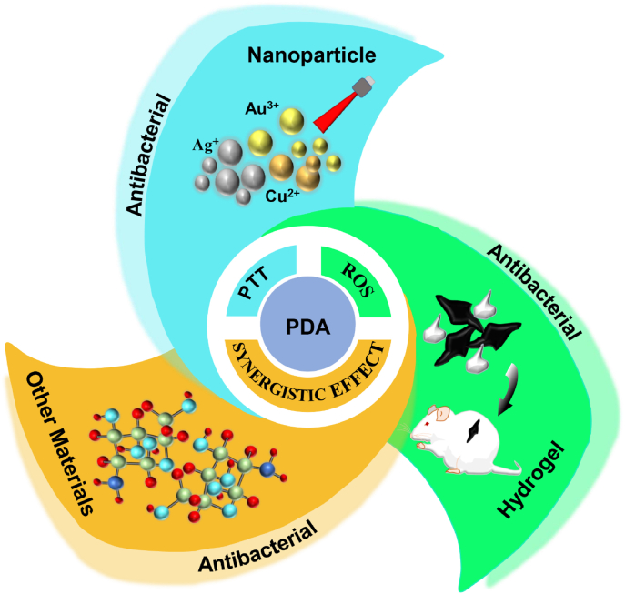
Antibiotic resistance is one of the major causes of morbidity and mortality, triggered by the adhesion of microbes and to some extent the formation of biofilms. This condition has been quite challenging in the health and industrial sector. Conditions and processes required to foil these infectious and resistance are of much concern. The synthesis of PDA material, inspired by the Mytilus edulis foot protein (MEFP)5 possesses unique characteristics that allow for, adhesion, photothermal therapy, synergistic effects with other materials, biocompatibility process, etc. Therefore, their usage holds great potential for dealing with both the infectious nature and the antibiotic resistance processes. Hence, this review provides an overview of the mechanism involved in accomplishing and eradicating bacteria, the recently harnessed antibacterial effect of the PDA through other properties they possess, a way forward in tapping the benefit embedded in the PDA, and the future perspective.

Keywords: Antibacterial activity; Antibiotic resistance; Biofilm; Nanoparticle; Polydopamine.


Conflict of interest statement

All authors agree to be published. All authors declare that they have no known competing financial interests or personal relationships that could have appeared to influence the work reported in this paper.

Figures

Image 1
Graphical abstract
Fig. 1
Fig. 1
Overview of the antibacterial mechanism of PDA and fabrication of PDA nanocomposites.
Fig. 2
Fig. 2
(a) Diagrammatic representation of the preparation process for FTCS-PDA/BNC membrane. (b) SEM images of BNC and FTCS-PDA/BNC membrane. (c) IR Images of FTCS-PDA/BNC membrane following exposure to 1 ​kW ​m−2 and 9 ​kW ​m−2. (d) Changes in temperature for BNC and FTCS-PDA/BNC membrane with/without water on top. (e) Measurement of the photothermal disinfection activity. With permission, reprinted from Ref. [84]. Copyright 2021 Elsevier.
Fig. 3
Fig. 3
(a) Diagrammatic illustration of the fabrication process for PDA/CS/Ag NPs for catheter coating and SEM images of uncoated and PDA/CS/Ag NPs-coated catheter (b) Fluorescent images of adhered bacteria on uncoated, PDA/CS coated and PDA/CS/Ag NPs coated Ti surfaces following incubation in S. aureus. (c) S. aureus inhibition halo test (d) SEM images of adhered bacteria on the surfaces of uncoated and PDA/CS/Ag NPs-coated catheters. With permission, reprinted from Ref. [44]. Copyright 2020 Elsevier.
Fig. 4
Fig. 4
(a) SEM images of the surfaces of TS, TS-M, and TS-M/P/V scaffolds (b) SEM images of TS, TS-M, and TS-M/P/V scaffolds with adhered S. aureus ( ​× ​8000 magnification). (c) Live/dead fluorescent images of S. aureus ( ​× ​200 magnification and 500 ​μm scale bar) (d) Representation of the biomass of bacteria and biofilm component of TS, TS-M, and TS-M/P/V. With permission, reprinted from Ref. [48]. Copyright 2018 Science China Materials.
Fig. 5
Fig. 5
(a) Schematic representation of the coating preparation for dual antifouling and antimicrobial activity. (b) Live/dead fluorescent images of adhered E. coli on unmodified and modified TiO2 (c) Quantitative representation of the number of adsorbed and dead E. coli on the unmodified and modified TiO2 substrate. (d) Plate-counting assay of S. epidermidis from catheters unmodified and those modified with r-pDA-40 and r-pDA-40-SBAA. With permission, reprinted from Ref. [91]. Copyright 2018 American Chemical Society.
Fig. 6
Fig. 6
(a) Diagrammatic illustration of the process involved in eliminating from Ti-M/I/RGD implant, already-established S. aureus's biofilm. (b) Acid etched Ti, Ti-M SEM images. (c) Images of S. aureus colonies following different treatment Ti, Ti-M, Ti-M/RGD, and Ti-M/I/RGD with the anti-biofilm efficiency. (d) SEM images of the morphology of bacteria treated with Ti, Ti ​+ ​NIR, Ti-M ​+ ​NIR (50 ​°C), Ti-M/RGD ​+ ​NIR (50 ​°C), Ti-M/I/RGD ​+ ​NIR (37 ​°C), and Ti-M/I/RGD ​+ ​NIR (50 ​°C), and (e) the images of the live/dead staining assay for the S. aureus. With permission, reprinted from Ref. [104]. Copyright 2019 Elsevier.
Fig. 7
Fig. 7
(a) Schematic representation of the combined activity of photothermal therapy and catalysis for antibacterial effect. (b) Diagrammatic representation of the synthesis of HAp, Au-HAp & PDA@Au-HAp with their respective SEM for characterization. (c) Changes in temperature with the infrared thermography for HAp, Au-HAp & PDA@Au-HAp under 808 ​nm laser irradiation (d) Images of bacteria colonies and antibacterial rates of E. coli and S. aureus following treatment with H2O2, HAp, Au-HAp, PDA@Au-HAp & PDA@Au-HAp ​+ ​H2O2. (e) Examination of the S. aureus infected wound healing after exposure to HAp, Au-HAp, PDA@Au-HAp ​+ ​NIR & PDA@Au-HAp ​+ ​H2O2 ​+ ​NIR. With permission, reprinted from Ref. [112]. Copyright 2018 Elsevier.
Fig. 8
Fig. 8
(a) Schematic illustration of the fabrication of PDA, melanin, Cu(II) loaded PDA, and Cu(II) loaded melanin nanoparticles for antibacterial activity. (b) SEM and TEM images of melanin, PDA, Cu(II) loaded melanin, and Cu(II) loaded PDA nanoparticles. (c) The E. coli and S. aureus inhibition efficiency of ampicillin, PDA, Cu(II) loaded PDA NPs, melanin, Cu(II) loaded melanin NPs and CuCl2, and (d) the corresponding live/dead fluorescent images. (e) Wound healing assessment for S. aureus infected wound, with PDA, Cu(II) loaded PDA nanoparticles within 14 days. With permission, reprinted from Ref. [114]. Copyright 2021 The Royal Society of Chemistry.
Fig. 9
Fig. 9
(a) Schematic illustration of the fabrication of the super hydrophilic coating of PDA with Ag+, with the antithrombotic and anti-infection. (b) Image of the PVC been coated with the PDA before and after with the coating morphology's SEM images. (c) Zone of inhibition for S. aureus and P. aeruginosa. (d) Live/Dead staining of adherent bacteria. (e) Adherent bacteria count for S. aureus and P. aeruginosa. With permission, reprinted from Ref. [125]. Copyright 2020 Elsevier.
Fig. 10
Fig. 10
(a) Schematic representation of the fabrication process of Ti-Nd-PDA-Fc. (b) SEM images of Ti–Nd, Ti-Nd-PDA, and Ti-Nd-PDA-Fc (c) Graphical representation of H2O2 generations for a specific time under a pH 7.4. (d) Images and quantification of MRSA and E. coli colonies treated with Ti–Nd, Ti-Nd-PDA, Ti-Nd-PDA-Fc, and Ti-Nd-PDA-Fc ​+ ​NIR under an acid and neutral environment (e) SEM images of the MRSA and E. coli morphology on substrates of Ti, Ti–Nd, Ti-Nd-PDA, Ti-Nd-PDA-Fc, and Ti-Nd-PDA-Fc ​+ ​NIR. (f) In vivo antibacterial test of MRSA infected wound, quantification and colonies of MRSA after treatment with Ti, Ti–Nd, Ti-Nd-PDA, Ti-Nd-PDA ​+ ​NIR, Ti-Nd-PDA-Fc, and Ti-Nd-PDA-Fc ​+ ​NIR. With permission, reprinted from Ref. [134]. Copyright 2020 American Chemical Society.
Fig. 11
Fig. 11
(a) Schematic representation of the synthesis of CP hydrogel and the synergistic antibacterial effect for periodontal therapy (b) SEM images of CP0, CP1, CP2, and CP3. (c) Photothermal changes, temperature based on NIR densities, and infrared thermography (d) Antibacterial assessment with CP3 alone with laser and CP3, CHX and CHX@CP3 with/without NIR irradiation against S. aureus. (e) Fluorescent staining and SEM images of live and dead S. aureus. With permission, reprinted from Ref. [161]. Copyright 2020 Elsevier.
Fig. 12
Fig. 12
(a) Schematic illustration of the preparation and application of XKP nanocomposite hydrogels. (b) SEM images of XK and XKP with high magnification image of XKP. (c) Photothermal effect and temperature change curves with photothermal images for XK containing varying concentrations of PDA. (d) Images of E. coli and S. aureus colonies following treatment with XK and XKP with the corresponding fluorescent images of the live/dead assay. (e) Representation of bacteria-infected skin wound with the size and area after treatment with PBS, XK, and XKP. With permission, reprinted from Ref. [160]. Copyright 2021 Elsevier.
Fig. 13
Fig. 13
(a) Schematic illustration of the fabrication of PDA/Alg/Fe3O4 (K3) beads. (b) HR-TEM, lattice fringes, and SAED images of PDA/Alg/Fe3O4 and (c) images of the prepared K1, K2, and K3 beads. (d) Bacteria inhibition efficiency based on the concentration of prepared K1, K2, and K3 beads after 12 ​h. (e) Variation in fluorescent of P·I for varying bacteria and a control. With permission, reprinted from Ref. [170]. Copyright 2019 The Royal Society of Chemistry.
Fig. 14
Fig. 14
(a) Schematic representation of the preparation and uses of the PEG-PDA (b) SEM images of PEG-PDA. (c) Images of the E. coli and MRSA colonies following 18 ​h of incubation with the SEM images representing the bacterial morphology. (d) Images of the E. coli and MRSA colonies following treatment PEG-PDA ​+ ​catalase and PEG-PDA with/without NIR (808 ​nm, 6 ​min) incubated for 1 ​h with the SEM images of bacterial morphology. (e) In vivo wound treatment with PEG and PEG-PDA, plating of fluid from the wound and the MRSA bacterial colonies count and histogram. With permission, reprinted from Ref. [177]. Copyright 2020 The Royal Society of Chemistry.
Fig. 15
Fig. 15
(a) Diagrammatic illustration of the synthesis of MM-PNAGAAu@ PDA for antibacterial activity. (b) Temperature changes for SMM-PNAGA and SMM-PNAGA ​+ ​varying concentration of Au@PDA under 808 ​nm (2 ​W ​cm−2) laser irradiation, a test with different laser powers, and the infrared thermography. (c) SEM images of E. coli and S. aureus following exposure to E/SMM-PNAGA ​+ ​NIR, and E/SMM-PNAGA-Au@PDA ​+ ​NIR. (d) In vivo wound healing activity assessment for S. aureus infected wound. With permission, reprinted from Ref. [184]. Copyright 2020 Elsevier.
Fig. 16
Fig. 16
(a) Optical and SEM images of GelNP-0, GelNP-025, GelNP-01 & GelNP-05. (b) Antibacterial assessment of GelNP-01 & GelNP-05 with the SEM images of adhered S. aureus. (c) Invitro skin disinfection test, with S. aureus infected skin, treated separately with saline solution and GelNP-05. (d) Cell viability and live/dead staining assay after incubation with 50 ​μM ​H2O2 (e) Wound healing assessment for S. aureus infected wound, diameter, and the wound area statistics. With permission, reprinted from Ref. [187]. Copyright 2021 Elsevier.
Fig. 17
Fig. 17
(a) Diagrammatic representation of PU/PDA (Cu)/PSBMA antibacterial and antifouling effect. (b) Antibacterial activity against E. coli and S. aureus and the fluorescent images of the live/dead assay following treatment with PU, PU/PDA, PU/PDA/PSBMA, PU/PDA (Cu), and PU/PDA (Cu)/PSBMA (c) SEM images of different magnification showing the anti-adhesive effect of PU, PU/PDA, PU/PDA/PSBMA, PU/PDA (Cu) and PU/PDA (Cu)/PSBMA (d) images S. aureus infected wound treated with PU, PU/PDA, PU/PDA/PSBMA, PU/PDA (Cu) and PU/PDA (Cu)/PSBMA (arrow indicates the pus), with LB plate containing S. aureus present in the pus after treatment with PU, PU/PDA, PU/PDA/PSBMA, PU/PDA (Cu) and PU/PDA (Cu)/PSBMA. With permission, reprinted from Ref. [190]. Copyright 2021 Elsevier.
Fig. 18
Fig. 18
(a) Diagrammatic illustration of antibacterial effect of DFT-C/ZnO-hydrogel. (b) Images and SEM images of DFT-hydrogel, TEM images of C/ZnO. (c)Photothermal curve with the temperature cooling profile and thermographic images of the various hydrogels. (d) Representation of the antibacterial activity of DFT-hydrogel, DFT-C/ZnO-hydrogel against S. aureus and E. coli, with/without 15 ​min of mixed light irradiation and SEM images of the bacteria. With permission, reprinted from Ref. [196]. Copyright 2019 WILEY-VCH Verlag GmbH & Co. KGaA, Weinheim.

Similar articles

Cited by

References

    1. Hall-Stoodley L., Costerton J.W., Stoodley P. Nat. Rev. Microbiol. 2004;2:95–108. - PubMed
    1. Zou Y., Zhang Y., Yu Q., Chen H. J. Mater. Sci. Technol. 2021;70:24–38.
    1. Amin Yavari S., Loozen L., Paganelli F.L., Bakhshandeh S., Lietaert K., Groot J.A., Fluit A.C., Boel C., Alblas J., Vogely H.C. ACS Appl. Mater. Interfaces. 2016;8:17080–17089. - PubMed
    1. Busscher H.J., van der Mei H.C., Subbiahdoss G., Jutte P.C., van den Dungen J.J., Zaat S.A., Schultz M.J., Grainger D.W. Sci. Transl. Med. 2012;4 153rv110. - PubMed
    1. Jia Z., Xiu P., Xiong P., Zhou W., Cheng Y., Wei S., Zheng Y., Xi T., Cai H., Liu Z. ACS Appl. Mater. Interfaces. 2016;8:28495–28510. - PubMed

LinkOut - more resources