. 2022 Jun 9:13:895501.
doi: 10.3389/fimmu.2022.895501. eCollection 2022.

Endothelin B Receptor Immunodynamics in Pulmonary Arterial Hypertension

Affiliations

Endothelin B Receptor Immunodynamics in Pulmonary Arterial Hypertension

Christoph Tabeling et al. Front Immunol. .

Abstract

Introduction: Inflammation is a major pathological feature of pulmonary arterial hypertension (PAH), particularly in the context of inflammatory conditions such as systemic sclerosis (SSc). The endothelin system and anti-endothelin A receptor (ETA) autoantibodies have been implicated in the pathogenesis of PAH, and endothelin receptor antagonists are routinely used treatments for PAH. However, immunological functions of the endothelin B receptor (ETB) remain obscure.

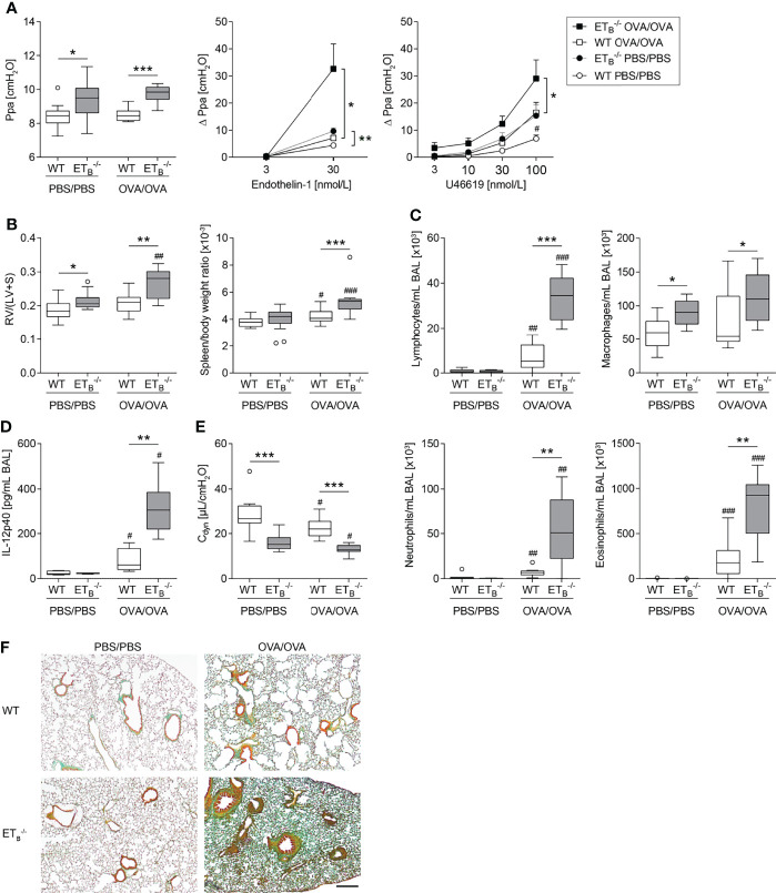
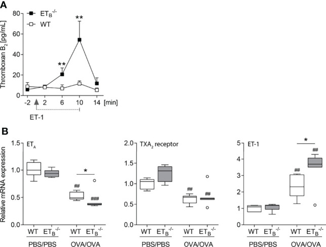
Methods: Serum levels of anti-ETB receptor autoantibodies were quantified in healthy donors and SSc patients with or without PAH. Age-dependent effects of overexpression of prepro-endothelin-1 or ETB deficiency on pulmonary inflammation and the cardiovascular system were studied in mice. Rescued ETB-deficient mice (ETB-/-) were used to prevent congenital Hirschsprung disease. The effects of pulmonary T-helper type 2 (Th2) inflammation on PAH-associated pathologies were analyzed in ETB-/- mice. Pulmonary vascular hemodynamics were investigated in isolated perfused mouse lungs. Hearts were assessed for right ventricular hypertrophy. Pulmonary inflammation and collagen deposition were assessed via lung microscopy and bronchoalveolar lavage fluid analyses.

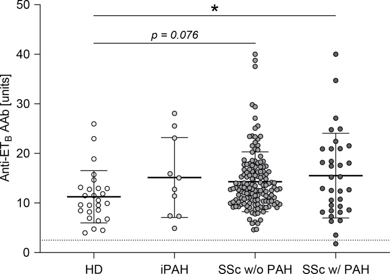
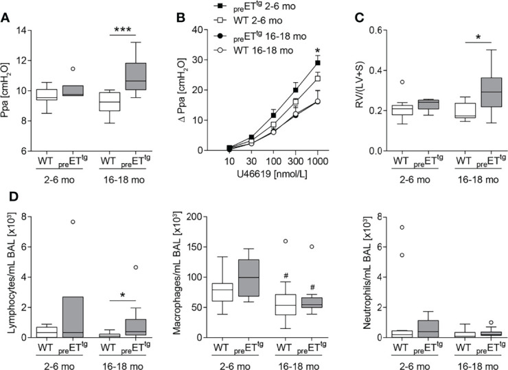
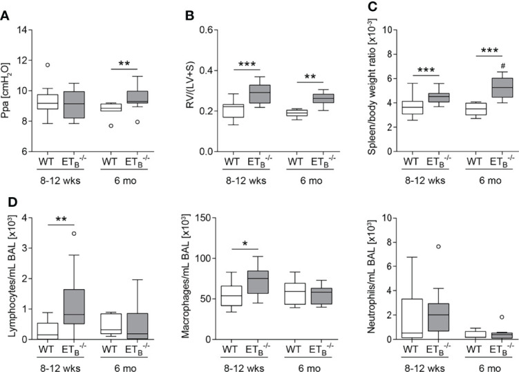
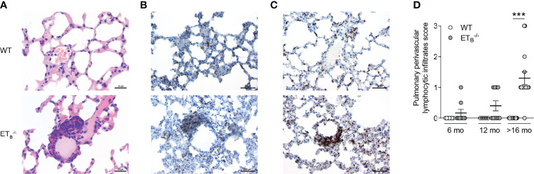
Results: Anti-ETB autoantibody levels were elevated in patients with PAH secondary to SSc. Both overexpression of prepro-endothelin-1 and rescued ETB deficiency led to pulmonary hypertension, pulmonary vascular hyperresponsiveness, and right ventricular hypertrophy with accompanying lymphocytic alveolitis. Marked perivascular lymphocytic infiltrates were exclusively found in ETB-/- mice. Following induction of pulmonary Th2 inflammation, PAH-associated pathologies and perivascular collagen deposition were aggravated in ETB-/- mice.

Conclusion: This study provides evidence for an anti-inflammatory role of ETB. ETB seems to have protective effects on Th2-evoked pathologies of the cardiovascular system. Anti-ETB autoantibodies may modulate ETB-mediated immune homeostasis.

Keywords: Th2 inflammation; autoantibody; endothelin B receptor; pulmonary arterial hypertension; pulmonary vascular hyperresponsiveness; systemic sclerosis.


Conflict of interest statement

CT received funding for research from Deutsche Gesellschaft für Pneumologie, Bayer HealthCare, Boehringer Ingelheim, and for lectures from Actelion Pharmaceuticals, Boehringer Ingelheim. HH is CEO of CellTrend GmbH, Luckenwalde, Germany. MW received funding for research from Deutsche Forschungsgemeinschaft, Bundesministerium für Bildung und Forschung, Deutsche Gesellschaft für Pneumologie, European Respiratory Society, Marie Curie Foundation, Else Kröner Fresenius Stiftung, CAPNETZ STIFTUNG, International Max Planck Research School, Actelion, Bayer Health Care, Biotest AG, Boehringer Ingelheim, NOXXON Pharma, Pantherna, Quark Pharma, Silence Therapeutics, Vaxxilon, and for lectures and advisory from Actelion, Alexion, Aptarion, Astra Zeneca, Bayer Health Care, Berlin Chemie, Biotest, Boehringer Ingelheim, Chiesi, Glaxo Smith Kline, Insmed, Novartis, Teva and Vaxxilon. The remaining authors declare that the research was conducted in the absence of any commercial or financial relationships that could be construed as a potential conflict of interest.

Figures

Figure 1
Figure 1
Anti-ETB autoantibody serum levels were elevated in patients with PAH secondary to SSc. Serum levels of anti-endothelin B receptor (ETB) autoantibodies (AAb) were quantified in healthy donors (HD), patients with idiopathic pulmonary arterial hypertension (iPAH), and systemic sclerosis (SSc) patients (+/- interstitial lung disease) with or without PAH. Data are expressed as single values with mean ± SD; N = 26 (HD) or N = 10 (iPAH) or N = 143 (SSc w/o PAH) or N = 34 (SSc w/PAH). The dotted line indicates the lower detection limit of the ELISA. *p < 0.05 (one-way ANOVA and Dunnett’s multiple comparisons test).
Figure 2
Figure 2
Prepro-endothelin-1 overexpression was age-dependently associated with increased pulmonary arterial pressure, vascular hyperresponsiveness, right ventricular hypertrophy, and increased number of lymphocytes in bronchoalveolar lavage. Lungs and hearts of 2- to 6-month (mo)-old or 16- to 18-mo-old prepro-endothelin-1 overexpressing (preETtg) mice and corresponding wild-type (WT) mice were prepared, or bronchoalveolar lavage (BAL) was performed. (A) In isolated perfused and ventilated lungs, under basal conditions, 16- to 18-mo-old preETtg showed a higher pulmonary arterial pressure (Ppa) compared to WT mice of the same age. (B) Pulmonary vascular responsiveness to intravascular application of the thromboxane receptor agonist U46619 was increased in 2- to 6-mo-old preETtg mice compared to WT controls of the same age. Data (Δ Ppa) represent the difference between the highest pressure response to U46619 and the basal Ppa. (C) Fulton index [quotient of right ventricle (RV) and left ventricle (LV) including septum (S)] determined after weighing the cardiac compartments was higher in 16- to 18-mo-old preETtg compared to WT mice of the same age. (D) Analysis of differentially quantified leukocytes in BAL showed increased number of lymphocytes in 16- to 18-mo-old preETtg compared to WT mice of the same age, whereas macrophages decreased with age, independent of prepro-ET-1 overexpression. In (A, C, D), data are represented as box plots depicting median, quartiles, and ranges excluding outliers (open circles), and analyzed using Mann–Whitney U test. # indicates significant difference between 16- to 18-mo-old vs. 2- to 6-mo-old groups, * indicates significant difference between preETtg vs. corresponding WT group (as indicated). In (B), values are given as mean and SEM, and analyzed using two-way repeated measures ANOVA, followed by a single Mann–Whitney U test between values of preETtg and WT mice of the same age at the highest dose of U46619 (*). In (A–C), N = 5–12; in (D), N = 7–17. */# p < 0.05, ***p < 0.001.
Figure 3
Figure 3
ETB deficiency was age-dependently associated with increased pulmonary arterial pressure, right ventricular hypertrophy, splenomegaly, and increased number of lymphocytes in bronchoalveolar lavage. Lungs, hearts, and spleens of 8- to 12-week (wk)-old and 6-mo-old rescued endothelin B receptor-deficient (ETB -/-) and corresponding wild-type (WT) mice were removed, or bronchoalveolar lavage (BAL) was performed. (A) In isolated perfused and ventilated lungs, under basal conditions, pulmonary arterial pressure (Ppa) was increased in 6-mo-old ETB -/- compared to WT mice of the same age. (B) Fulton index [ratio of right ventricle (RV) and left ventricle (LV) including septum (S)] determined after weighing the cardiac compartments was higher in ETB -/- compared to WT mice. (C) Determination of spleen weight related to body weight revealed splenomegaly in ETB -/- mice. (D) Analysis of differentially quantified leukocytes in BAL revealed increased number of lymphocytes and macrophages in BAL from 8- to 12-wk-old ETB -/- vs. WT mice of the same age. Data are represented as box plots depicting median, quartiles, and ranges excluding outliers (open circles). In (A–C), N = 7–28; in (D), N = 7–17. # indicates significant difference in the 6-mo-old vs. the corresponding 8- to 12-wk-old group, * indicates significant difference between ETB -/- vs. the corresponding WT group. */# p < 0.05, **p < 0.01, ***p < 0.001 (Mann–Whitney U test).
Figure 4
Figure 4
ETB deficiency was associated with peripheral perivascular lymphocytic infiltrates in the lung. Lungs of 6-, 12-, and >16-mo-old rescued endothelin B receptor-deficient (ETB -/-) and the corresponding wild-type (WT) mice were assessed histologically following hematoxylin and eosin (H&E) stain (A) or immunohistochemical stains for CD45R/B220 (B cells; B) or CD3 (T cells; C). The scale bars represent 20 µm (A) or 50 µm (B, C). Representative images of ≥12-mo-old mice are shown; N = 20–25 per group (A) or N = 3–5 per group (B, C). (D) H&E-stained lung sections were analyzed and scored (0, no peripheral perivascular infiltrates; 1, mild; 2 moderate; 3, pronounced) by an independent board-certified pathologist, blinded to the study groups. Data are expressed as single values with mean ± SEM; N = 7–9 (6-mo-old group), N = 5–10 (12-mo-old group), or N = 15 (>16-mo-old group). ***p < 0.001 (Mann–Whitney U test).
Figure 5
Figure 5
ETB deficiency aggravated Th2-mediated vascular pathologies and inflammation in the lung. Rescued endothelin B receptor-deficient (ETB -/-) and corresponding wild-type (WT) mice were systemically sensitized with ovalbumin (OVA) (or PBS as control) and repeatedly exposed to aerosolized OVA (OVA/OVA) or PBS (PBS/PBS). Forty-eight hours after the last challenge, lungs, hearts, and spleens of 12-wk-old mice were harvested, or bronchoalveolar lavage (BAL) was performed. (A) In isolated perfused and ventilated lungs, pulmonary arterial pressure (Ppa) was measured under basal conditions, and pulmonary vascular responsiveness to increasing concentrations of endothelin-1 or thromboxane receptor agonist U46619 was determined. Data (Δ Ppa) represent the difference between the highest pressure response to the respective stimulus and the basal Ppa. (B) Fulton index [quotient of right ventricle (RV) and left ventricle (LV) including septum (S)] was determined after weighing the cardiac compartments (left) and spleen weight was determined and related to body weight (right). (C) Leukocytes were differentially quantified in BAL. (D) IL-12p40 was determined in BAL (lower detection limit was 0.54 pg/mL). (E) In isolated perfused and ventilated mouse lungs, dynamic lung compliance (Cdyn) was measured. (F) Lung tissue sections were stained with Masson–Goldner trichrome that revealed more pronounced pulmonary collagen deposition in ETB -/- than WT mice after OVA/OVA treatment. The scale bar represents 100 µm and is valid for all photomicrographs. Representative images (N = 7 per group) are shown. In (A left, B–E), data are represented as box plots depicting median, quartiles, and ranges excluding outliers (open circles), and analyzed using Mann–Whitney U test. # indicates significant difference between OVA/OVA vs. the corresponding PBS/PBS group, * indicates significant difference between ETB -/- vs. the corresponding WT group. In (A middle-right), values are given as mean and SEM, and analyzed using two-way repeated measures ANOVA (*). In (A right), additional Mann–Whitney U test was performed comparing values of ETB -/- and WT mice treated with PBS at the highest dose of U46619 (#). N = 5–14 (A–C, E) or N = 3–6 (D). */# p < 0.05, **/## p < 0.01, ***/### p < 0.001.
Figure 6
Figure 6
ET-1-mediated thromboxane release is increased in ETB -/- mice. (A) In isolated perfused lungs of 10- to 12-wk-old rescued endothelin B receptor-deficient (ETB -/-) and corresponding wild-type (WT) mice, perfusate samples were collected 2 min before application of 100 nmol/L ET-1 (for 10 min), and 2, 6, 10, and 14 min after the start of ET-1 application, and TXB2 levels were determined. The detection limit was 7.8 pg/ml. Values are given as mean and SEM (N = 7), and analyzed using Mann–Whitney U tests at each time point comparing both groups. (B) ETB -/- and corresponding WT mice were systemically sensitized with OVA (or PBS as control) and repeatedly exposed to aerosolized OVA (OVA/OVA) or PBS (PBS/PBS). Forty-eight hours after the last inhalative OVA challenge, lungs of 15- to 17-wk-old mice were isolated for mRNA expression analyses by quantitative PCR. Relative quantification of mRNA was performed using the comparative Ct method. Data are represented as box plots depicting median, quartiles, and ranges excluding outliers (open circles), and analyzed using Mann–Whitney U test; N = 67 per group. # indicates significant difference between OVA/OVA vs. the corresponding PBS/PBS group, * indicates significant difference between ETB -/- vs. the corresponding WT group. *p < 0.05, **/## p < 0.01, ### p < 0.001.

References

    1. Gaine S. Pulmonary Hypertension. JAMA (2000) 284(24):3160–8. doi: 10.1001/jama.284.24.3160 - DOI - PubMed
    1. Price LC, Wort SJ, Perros F, Dorfmüller P, Huertas A, Montani D, et al. . Inflammation in Pulmonary Arterial Hypertension. Chest (2012) 141(1):210–21. doi: 10.1378/chest.11-0793 - DOI - PubMed
    1. Hassoun PM, Mouthon L, Barberà JA, Eddahibi S, Flores SC, Grimminger F, et al. . Inflammation, Growth Factors, and Pulmonary Vascular Remodeling. J Am Coll Cardiol (2009) 54(1 Suppl):S10–9. doi: 10.1016/j.jacc.2009.04.006 - DOI - PubMed
    1. Huertas A, Tu L, Humbert M, Guignabert C. Chronic Inflammation Within the Vascular Wall in Pulmonary Arterial Hypertension: More Than a Spectator. Cardiovasc Res (2020) 116(5):885–93. doi: 10.1093/cvr/cvz308 - DOI - PubMed
    1. Kherbeck N, Tamby MC, Bussone G, Dib H, Perros F, Humbert M, et al. . The Role of Inflammation and Autoimmunity in the Pathophysiology of Pulmonary Arterial Hypertension. Clin Rev Allergy Immunol (2013) 44(1):31–8. doi: 10.1007/s12016-011-8265-z - DOI - PubMed

Publication types

MeSH terms

LinkOut - more resources