. 2022 Jun 10:13:916512.
doi: 10.3389/fimmu.2022.916512. eCollection 2022.

Heparanase Is a Putative Mediator of Endothelial Glycocalyx Damage in COVID-19 - A Proof-of-Concept Study

Affiliations

Heparanase Is a Putative Mediator of Endothelial Glycocalyx Damage in COVID-19 - A Proof-of-Concept Study

Carolin Christina Drost et al. Front Immunol. .

Abstract

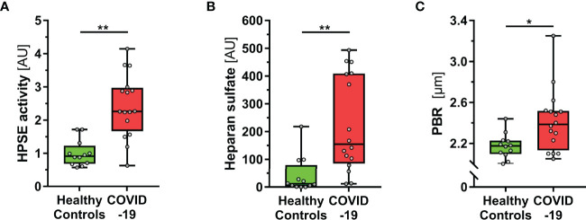
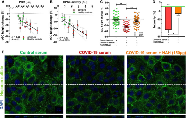
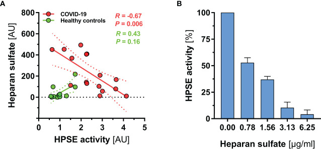
Coronavirus disease 2019 (COVID-19) is a systemic disease associated with injury (thinning) of the endothelial glycocalyx (eGC), a protective layer on the vascular endothelium. The aim of this translational study was to investigate the role of the eGC-degrading enzyme heparanase (HPSE), which is known to play a central role in the destruction of the eGC in bacterial sepsis. Excess activity of HPSE in plasma from COVID-19 patients correlated with several markers of eGC damage and perfused boundary region (PBR, an inverse estimate of glycocalyx dimensions of vessels with a diameter 4-25 µm). In a series of translational experiments, we demonstrate that the changes in eGC thickness of cultured cells exposed to COVID-19 serum correlated closely with HPSE activity in concordant plasma samples (R = 0.82, P = 0.003). Inhibition of HPSE by a nonanticoagulant heparin fragment prevented eGC injury in response to COVID-19 serum, as shown by atomic force microscopy and immunofluorescence imaging. Our results suggest that the protective effect of heparin in COVID-19 may be due to an eGC-protective off-target effect.

Keywords: COVID-19; endothelial glycocalyx (EG); heparanase (HPSE); heparin; videomicroscopy.


Conflict of interest statement

The authors declare that the research was conducted in the absence of any commercial or financial relationships that could be construed as a potential conflict of interest.

Figures

Figure 1
Figure 1
COVID-19 patients show elevated HPSE activity, and damaged eGC in vivo. (A-C) Boxplots showing (A) heparanase (HPSE) activity, (B) heparan sulfate (HS) and (C) perfused boundary region (PBR; an inverse estimate of the sublingual endothelial glycocalyx thickness) in healthy subjects (n = 12) and COVID-19 patients at the ICU (n = 16). Differences between groups were calculated by Mann-Whitney U test. *p < 0.05; **p < 0.001.
Figure 2
Figure 2
HPSE is a putative mediator of eGC damage in COVID-19. (A-C) Sera from a randomly selected subgroup of 5 healthy controls and 6 COVID-19 patients were sterile-filtered and incubated (5%) on the human umbilical vein endothelial cell line EA.hy926 for 60 min. Endothelial glycocalyx (eGC) thickness was assessed by atomic force microscopy (AFM) using a dedicated nano-indentation protocol. Scatter dot plot showing the association between AFM-derived eGC (in vitro) decline and corresponding (A) PBR-values (in vivo) and (B) HPSE activity for the individuals from the subgroup. Each dot represents the mean ± SEM (standard error of mean) of two independent AFM experiments (consisting of ≥ 4 indentation curves in each of ≥ 8 different cells) for each individual serum. Incubation without human serum served as control. Correlation was assessed by Spearman correlation coefficient. (C) Dot plots from three independent AFM experiments (pooled serum from subgroups) showing values with mean ± SEM. Each dot represents the mean of ≥ 4 indentation curves per cell. Heparanase was blocked by N-desulfated re-N-acetylated heparin (NAH; 150 µg). Differences between groups were calculated with nested ANOVA and Tukey’s post-hoc test. Intensity analysis of heparan sulfate-stained EA.hy926 cells (D) and representative immunofluorescence images (E) after treatment with 5% control serum or COVID-19 serum ± NAH (150 µg) for 60 min. Values are normalized to control serum (zero line) and differences between groups were assessed with nested ANOVA and Tukey’s post-hoc test. Data are presented as mean ± SEM. *p < 0.05; **p < 0.001.
Figure 3
Figure 3
HS fragments in severe COVID-19 may partially block HPSE activity. (A) Scatter dot plot showing regression slopes of HPSE activity vs. HS plasma concentration in healthy subjects (n = 12) and COVID-19 patients (n = 16), respectively. (B) Bar charts showing percentage decrease of HPSE activity with increasing amounts of HS (isolated from bovine kidney) in three independent experiments. For this experiment, recombinant human HPSE was used in a concentration of 150 ng/ml. Data are presented as mean ± SEM.

Similar articles

Cited by

References

    1. Smadja DM, Mentzer SJ, Fontenay M, Laffan MA, Ackermann M, Helms J, et al. . Covid-19 Is a Systemic Vascular Hemopathy: Insight for Mechanistic and Clinical Aspects. Angiogenesis (2021) 24(4):755–88. doi: 10.1007/s10456-021-09805-6 - DOI - PMC - PubMed
    1. Pons S, Fodil S, Azoulay E, Zafrani L. The Vascular Endothelium: The Cornerstone of Organ Dysfunction in Severe Sars-Cov-2 Infection. Crit Care (2020) 24(1):353. doi: 10.1186/s13054-020-03062-7 - DOI - PMC - PubMed
    1. Emadi-Baygi M, Ehsanifard M, Afrashtehpour N, Norouzi M, Joz-Abbasalian Z. Corona Virus Disease 2019 (Covid-19) as a System-Level Infectious Disease With Distinct Sex Disparities. Front Immunol (2021) 12:778913. doi: 10.3389/fimmu.2021.778913 - DOI - PMC - PubMed
    1. Rovas A, Osiaevi I, Buscher K, Sackarnd J, Tepasse PR, Fobker M, et al. . Microvascular Dysfunction in Covid-19: The Mystic Study. Angiogenesis (2021) 24(1):145–57. doi: 10.1007/s10456-020-09753-7 - DOI - PMC - PubMed
    1. Reitsma S, Slaaf DW, Vink H, van Zandvoort MA, oude Egbrink MG. The Endothelial Glycocalyx: Composition, Functions, and Visualization. Pflugers Arch (2007) 454(3):345–59. doi: 10.1007/s00424-007-0212-8 - DOI - PMC - PubMed

Publication types