. 2022 Nov 7;43(42):4496-4511.
doi: 10.1093/eurheartj/ehac337.

A circular RNA derived from the insulin receptor locus protects against doxorubicin-induced cardiotoxicity

Affiliations

A circular RNA derived from the insulin receptor locus protects against doxorubicin-induced cardiotoxicity

Dongchao Lu et al. Eur Heart J. .

Abstract

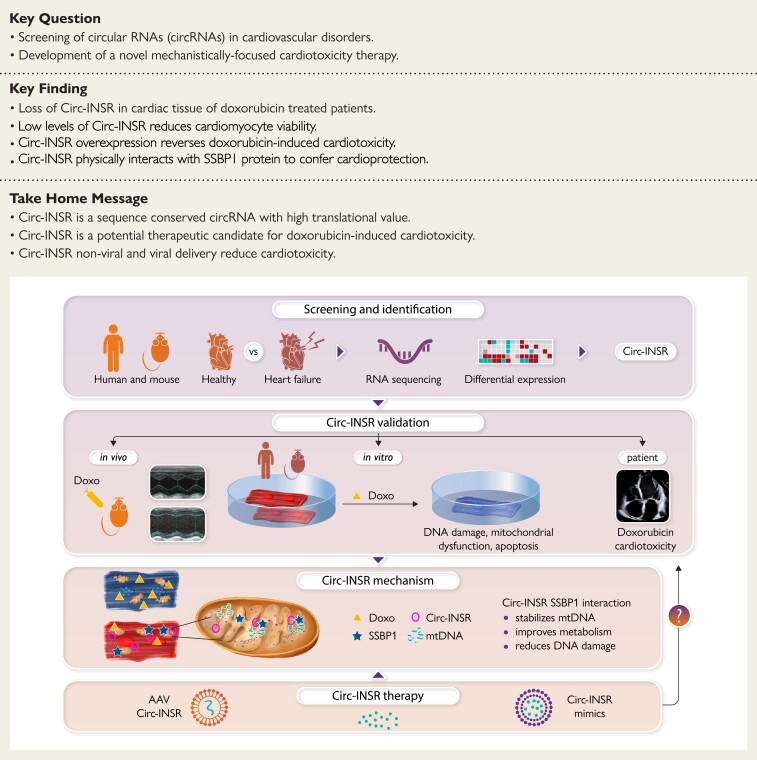
Aims: Cardiotoxicity leading to heart failure (HF) is a growing problem in many cancer survivors. As specific treatment strategies are not available, RNA discovery pipelines were employed and a new and powerful circular RNA (circRNA)-based therapy was developed for the treatment of doxorubicin-induced HF.

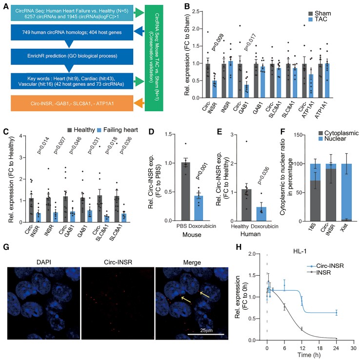
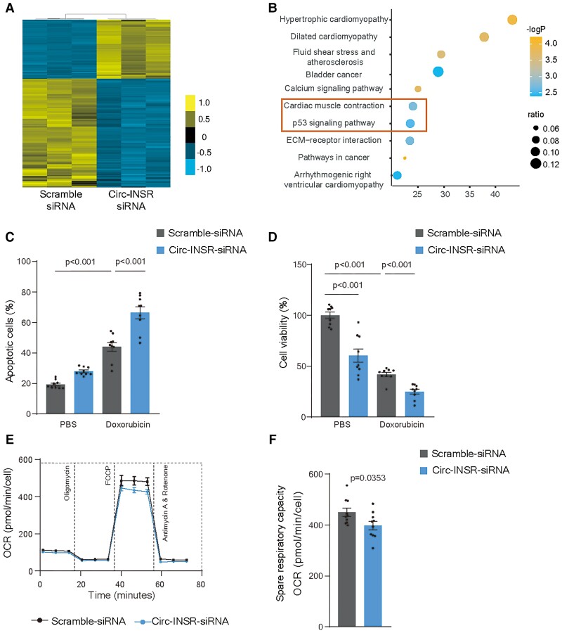
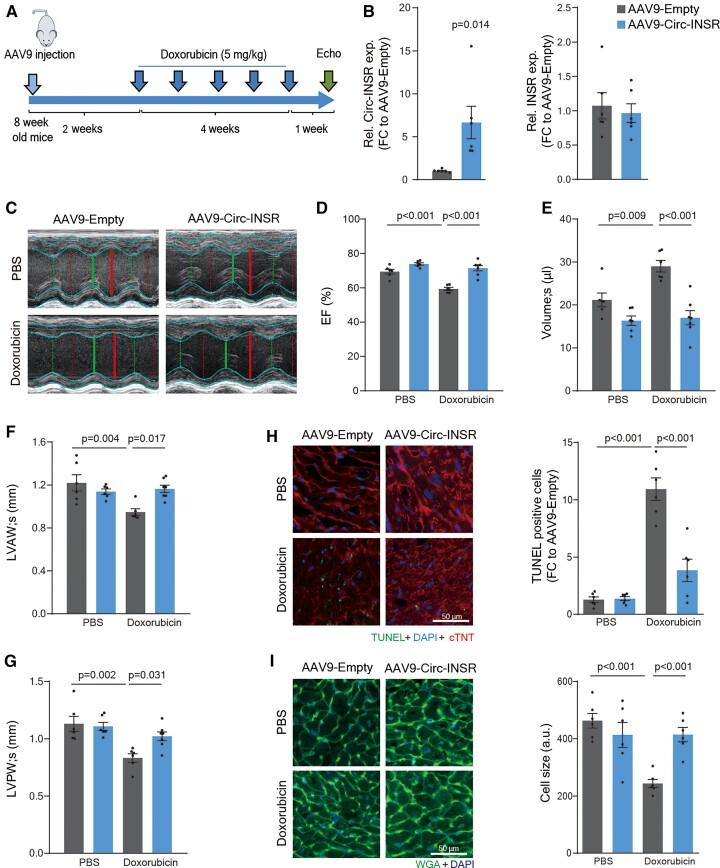
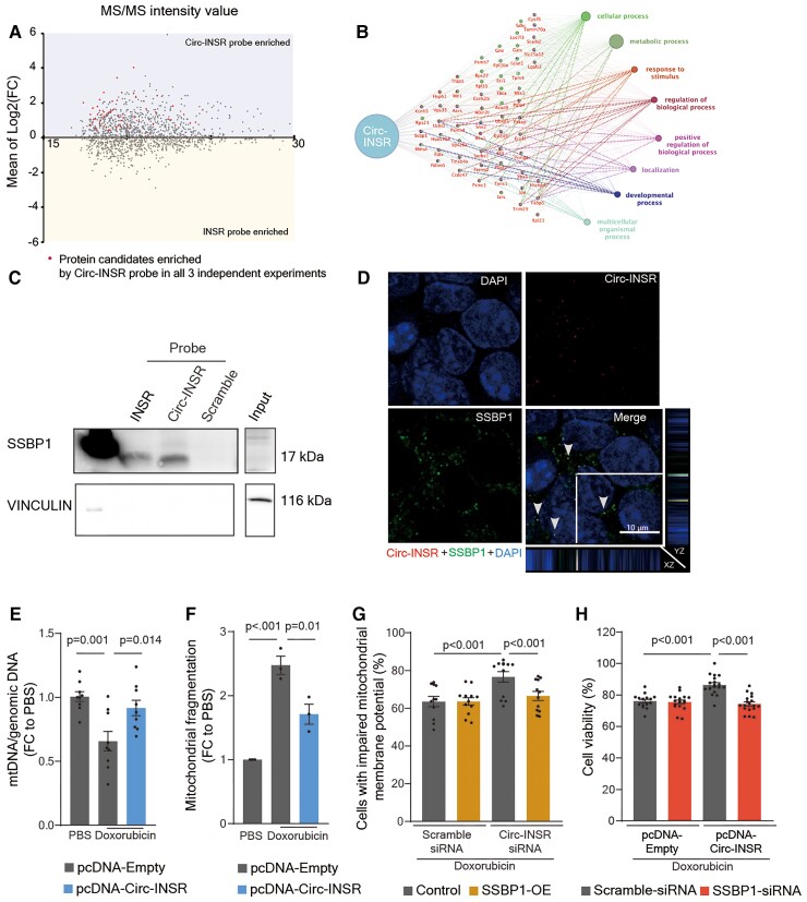
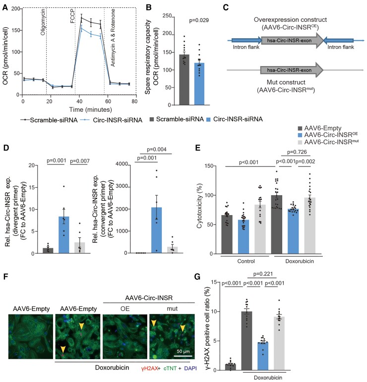
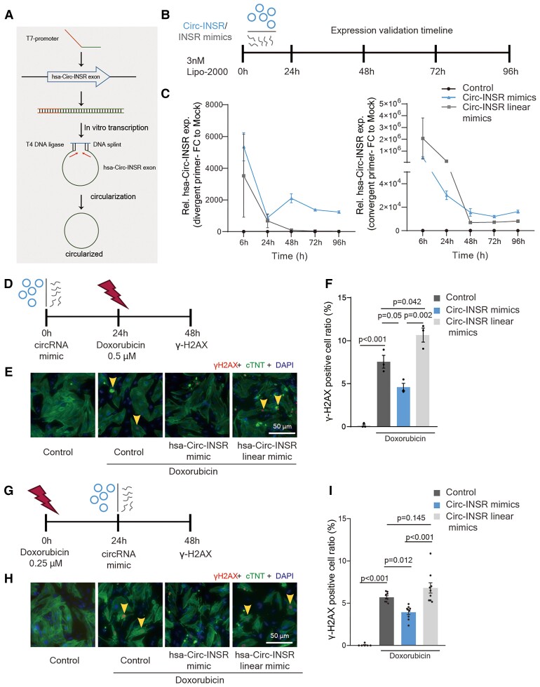
Methods and results: The circRNA sequencing was applied and the highly species-conserved circRNA insulin receptor (Circ-INSR) was identified, which participates in HF processes, including those provoked by cardiotoxic anti-cancer treatments. Chemotherapy-provoked cardiotoxicity leads to the down-regulation of Circ-INSR in rodents and patients, which mechanistically contributes to cardiomyocyte cell death, cardiac dysfunction, and mitochondrial damage. In contrast, Circ-INSR overexpression prevented doxorubicin-mediated cardiotoxicity in both rodent and human cardiomyocytes in vitro and in a mouse model of chronic doxorubicin cardiotoxicity. Breast cancer type 1 susceptibility protein (Brca1) was identified as a regulator of Circ-INSR expression. Detailed transcriptomic and proteomic analyses revealed that Circ-INSR regulates apoptotic and metabolic pathways in cardiomyocytes. Circ-INSR physically interacts with the single-stranded DNA-binding protein (SSBP1) mediating its cardioprotective effects under doxorubicin stress. Importantly, in vitro transcribed and circularized Circ-INSR mimics also protected against doxorubicin-induced cardiotoxicity.

Conclusion: Circ-INSR is a highly conserved non-coding RNA which is down-regulated during cardiotoxicity and cardiac remodelling. Adeno-associated virus and circRNA mimics-based Circ-INSR overexpression prevent and reverse doxorubicin-mediated cardiomyocyte death and improve cardiac function. The results of this study highlight a novel and translationally important Circ-INSR-based therapeutic approach for doxorubicin-induced cardiac dysfunction.

Keywords: Heart failure • Circular RNA • Doxorubicin cardiotoxicity • AAVtherapy • Mitochondrial metabolism • Anti-cancer treatment.


Conflict of interest statement

Conflict of interest: T.T. is a founder and shareholder of Cardior Pharmaceuticals GmbH (outside the topic of this paper). D.C.L., C.B., and T.T. have filed and partly licensed patents for ncRNAs including one patent for Circ-INSR.

Figures

Structured Graphical Abstract
Circular RNA therapy for cardioprotection. AAV, adeno-associated virus; INSR, insulin receptor; SSBP1, single-stranded DNA-binding protein.
Figure 1
Figure 1
Identification of Circ-INSR from failing hearts and validation in various heart failure models. (A) Pipeline to select candidates from RNA-Seq data. (B) Relative expression of circRNA and their linear host genes (Circ-INSR, INSR, Circ-GAB1, GAB1, Circ-SLC8A1, SLC8A1, Circ-ATP1A1, and ATP1A1) in TAC model at 13 weeks (n = 8 mice/group). (C) Relative expression of circRNA and their linear host genes (Circ-INSR, INSR, Circ-GAB1, GAB1, Circ-SLC8A1, and SLC8A1) in failing heart (n = 6 patients) compared with healthy (n = 12), expression of Circ-ATP1A1 was not detectable in the human samples. (D) Relative expression of Circ-INSR in mouse hearts treated with doxorubicin (n = 6 per group). (E) Relative expression of Circ-INSR in male patient heart with doxorubicin-induced heart failure (n = 4) compared with healthy (n = 10; Mann–Whitney U test was performed). (F) Circ-INSR expression in neonatal rat cardiomyocytes represented after normalization to the respective subcellular fraction (18S applied for normalization of cytoplasmic fraction, Xist applied for normalization of nuclear fraction, n = 6 per group from duplicates of three independent experiments). (G) RNA fluorescence in situ hybridization (RNA-FISH) of Circ-INSR in HL-1 cells (scale bar, 25 μm). Thin yellow arrows indicate cytoplasmic localization of the Circ-INSR as observed in RNA-FISH experiment. (H) Relative expression of Circ-INSR and INSR in HL-1 cells treated with Actinomycin D (n = 6 per timepoint). All quantitative data are presented as mean ± standard error mean, and an unpaired two-tailed t-test or Mann–Whitney U test was performed to calculate significance between two groups. P < 0.05 was considered as statistically significant. exp., expression; FC, fold change; GO, gene ontology; NRCM, neonatal rat cardiomyocyte; TAC, transverse aortic constriction; Rel., relative.
Figure 2
Figure 2
Down-regulation of Circ-INSR reduces cardiomyocytes survival and disrupts metabolic activity. (A) Heat map of RNA-Seq data for selected genes from the HL-1 cells treated with Circ-INSR-siRNA compared with Scramble siRNA (blue indicates down-regulated genes, yellow indicates up-regulated genes). (B) KEGG analysis of the dysregulated gene pathways from the RNA sequencing data. Orange box in the bubble plot highlights the most prominently regulated cellular processes after Circ-INSR knockdown. (C) Annexin-V and 7-AAD staining of Circ-INSR-siRNA transfected HL-1 cells with/without doxorubicin treatment (0.25 μM 48 h, n = 9 wells per group from triplicates of three independent experiments, one-way analysis of variance was performed within Scramble siRNA, Scramble siRNA + Doxorubicin, and Circ-INSR-siRNA + Doxorubicin). (D) Percentage of viable cells (WST-1 assay) in Circ-INSR-siRNA treated HL-1 cells compared with Scramble siRNA with/without doxorubicin treatment (0.25 μM 48 h, n = 9 wells per group from triplicates of three independent experiments, one-way analysis of variance was performed within Scramble siRNA, Scramble siRNA + Doxorubicin, and Circ-INSR-siRNA + Doxorubicin, unpaired two-tailed t-test was performed between Scramble siRNA and Circ-INSR-siRNA). (E) Mean oxygen consumption rate at various time points of Mito stress assay (n = 10 wells per group from one experiment). (F) Spare respiratory capacity of neonatal mouse cardiomyocytes treated with Circ-INSR-siRNA compared with Scramble siRNA (n = 10 wells per group from one experiment). All quantitative data are presented as mean ± standard error mean, and an unpaired two-tailed t-test was performed to calculate significance between two groups, and one-way analysis of variance with post hoc Tukey test was used to calculate significance between ≥3 groups wherever required. P < 0.05 was considered as statistically significant. NMCM, neonatal mouse cardiomyocyte; OCR, oxygen consumption rate.
Figure 3
Figure 3
Circ-INSR overexpression alleviates doxorubicin-induced metabolic disorder and cardiomyocyte apoptosis. (A) Percentage of apoptotic cells quantified by Annexin-V and 7-AAD staining of pcDNA-Empty and pcDNA-Circ-INSR transfected HL-1 cells with/without doxorubicin (0.25 μM 48 h, n = 12/6/19/18 from three independent experiments, one-way analysis of variance was performed within pcDNA-Empty, pcDNA-Empty + Doxorubicin, and pcDNA-Circ-INSR + Doxorubicin). (B) Percentage of viable HL-1 cells (WST-1 assay) in pcDNA-Circ-INSR overexpression HL-1 cells compared with pcDNA-Empty control (0.25 μM 48 h, n = 12/8/8/10 from three independent experiments, one-way analysis of variance was performed within pcDNA-Empty, pcDNA-Empty + Doxorubicin, and pcDNA-Circ-INSR + Doxorubicin, unpaired two-tailed t-test was performed between pcDNA-Empty and pcDNA-Circ-INSR). (C) γ-H2AX (red) staining in nuclei (DAPI, blue) of neonatal rat cardiomyocytes (cTNT, green) transduced with either AAV6-Empty control or AAV6-Circ-INSR, in the presence or absence of doxorubicin. Yellow arrows denote DNA damage (0.25 μM 48 h, n = 8/6/8/8 from three independent experiments, one-way analysis of variance was performed within AAV6-Empty, AAV6-Empty + Doxorubicin, and AAV6-Circ-INSR + Doxorubicin). Representative images left; quantification right (scale bar, 50 μm). (D) TUNEL positive (green) staining in nuclei (DAPI, blue) of neonatal rat cardiomyocytes (cTNT, red) transduced with either AAV6-Empty control or AAV6-Circ-INSR, in the presence or absence of doxorubicin. Yellow arrows indicate apoptotic cells (0.25 μM 48 h, n = 6/5/6/6 from three independent experiments, one-way analysis of variance was performed within AAV6-Empty, AAV6-Empty + Doxorubicin, and AAV6-Circ-INSR + Doxorubicin); Representative images left; quantification right (scale bar, 50 μm). (E) Mean oxygen consumption rate at various time points of Mito Stress Assay (5 μM, 2 h, n = 17/16/16/16 wells from one experiment). (F and G) Basal respiration and spare respiratory capacity of NMCMs treated with AAV6-Circ-INSR compared with AAV6-Empty under basal conditions or doxorubicin treatment (5 μM, 2 h, n = 17/16/16/16 wells from one experiment; one-way analysis of variance (Bonferroni’s multiple comparison test) was performed within AAV6-Empty, AAV6-Empty + Doxorubicin and AAV6-Circ-INSR + Doxorubicin, unpaired two-tailed t-test was performed between AAV6-Empty and AAV6-Circ-INSR). All quantitative data are presented as mean ± standard error mean, and one-way analysis of variance with post hoc Tukey test or Bonferroni’s multiple comparison test was used to calculate significance between ≥3 groups wherever required. P < 0.05 was considered as statistically significant. FC, fold change; NRCM/NMCM, neonatal rat/mouse cardiomyocyte; OCR, oxygen consumption rate.
Figure 4
Figure 4
Circ-INSR therapy protects from doxorubicin-induced cardiotoxicity in vivo. (A) Schematic representation of the in vivo animal model of chronic doxorubicin-induced heart failure. (B) Relative expression of Circ-INSR and INSR in AAV9-Circ-INSR and AAV9-Empty heart tissue (n = 6 mice/group). (C) Representative figures of echocardiography (Echo) after AAV9-Circ-INSR therapy in presence or absence of doxorubicin cardiotoxicity. (DG) Ejection fraction, left ventricular end-systolic volume (volume; s), dimension (in mm) of the anterior wall of the left ventricle at systole (s), dimension (in mm) of the posterior wall of the left ventricle at systole (s) in AAV9-Circ-INSR and AAV9-Empty-treated mice with or without doxorubicin (n = 6/6/6/7 mice, one-way analysis of variance was performed within AAV9-Empty, AAV9-Empty + Doxorubicin, and AAV9-Circ-INSR + Doxorubicin). (H) TUNEL (green) co-staining with DAPI in nuclei (blue) and cTNT (red) in AAV9-Empty and AAV9-Circ-INSR heart sections with/without doxorubicin treatment; representative images left; quantification right (n = 6 mice/group, scale bar, 50 μm, one-way analysis of variance was performed within AAV9-Empty, AAV9-Empty + Doxorubicin, and AAV9-Circ-INSR + Doxorubicin). (I) WGA (green) staining in AAV9-Empty and AAV9-Circ-INSR heart sections with/without doxorubicin treatment. Blue staining indicates DAPI-stained nuclei. Representative images left; quantification right (n = 6 mice/group, scale bar, 50 μm, one-way analysis of variance was performed within AAV9-Empty, AAV9-Empty + Doxorubicin, and AAV9-Circ-INSR + Doxorubicin). All quantitative data are presented as mean ± standard error mean, and an unpaired two-tailed t-test was performed to calculate significance between two groups, and one-way analysis of variance with post hoc Tukey test was used to calculate significance between ≥3 groups wherever required. P < 0.05 was considered as statistically significant. a.u., arbitrary units; EF, ejection fraction; exp., expression; FC, fold change; LVAW, anterior wall of the left ventricle at systole; LVPW, posterior wall of the left ventricle at systole; Rel., relative.
Figure 5
Figure 5
Circ-INSR is regulated by Brca1. (A) Prediction of the upstream regulator of Circ-INSR using the RegRNA2.0 tool. (B) Relative expression of Brca1 in mouse heart tissue with or without doxorubicin treatment (n = 6 mice per group). (C) Relative expression of Brca1 in HL-1 cells treated with Brca1-siRNA compared with Scramble siRNA (n = 7, from two independent experiments). (D) Relative expression of Circ-INSR and INSR in HL-1 cells treated with Brca1-siRNA compared with Scramble siRNA (n = 7, from two independent experiments). (E) Relative expression of Circ-INSR and INSR in pcDNA-Circ-INSR transfected HL-1 cells co-treated with Brca1-siRNA compared with control transfected with pcDNA-Empty and Scramble siRNA (n = 7, from two independent experiments, one-way analysis of variance performed within Control, pcDNA-Circ-INSR + Scramble siRNA, and pcDNA-Circ-INSR + Brca1-siRNA). All quantitative data are presented as mean ± standard error mean, and an unpaired two-tailed t-test was performed to calculate significance between two groups and one-way analysis of variance with post hoc Tukey test was used to calculate significance between ≥3 groups wherever required. P < 0.05 was considered as statistically significant. exp., expression; FC, fold change; Rel., relative.
Figure 6
Figure 6
Circ-INSR regulates metabolism and cell viability via interaction with single-stranded DNA-binding protein 1 in cardiomyocytes. (A) MA plot showing the relationship between intensity value and fold change (log2FC) among the proteins. Only those proteins which were enriched by the Circ-INSR probe in all three individual experiments were labelled with red dots. (B) Interaction network laid out and visualized with ClueGO plugin of Cytoscape. Dots indicate the proteins interacting with Circ-INSR, and the respective colours indicate the interaction types. Each gene ontology pathway is depicted by a specific node colour. The size of the node represents the number of interacting proteins, which belong to the respective gene ontology pathway. (C) Western blot of single-stranded DNA-binding protein 1 levels in HL-1 cells after Circ-INSR and INSR RNA pulldown using biotin-labelled DNA probes. (D) RNA fluorescence in situ hybridization (RNA-FISH) of Circ-INSR (red) and co-staining with single-stranded DNA-binding protein 1 (green) and DAPI (blue) in HL-1 cells (scale bar, 10 μm); Z-stack view of the co-localized single-stranded DNA-binding protein 1 and Circ-INSR molecules are shown around the edge of the merge figure panel. Grey arrows highlight the yellow dots which indicate the co-localization of Circ-INSR and single-stranded DNA-binding protein 1 in the cytoplasm. (E) Mitochondrial DNA (mtDNA) copy number analysis of HL-1 cells treated with pcDNA-Circ-INSR overexpression plasmid and doxorubicin compared with pcDNA-Empty control (0.25 μM 48 h, n = 9 wells per group from triplicates of three independent experiments, one-way analysis of variance was performed). (F) Quantification of MitoTracker staining assay in HL-1 cells treated with pcDNA-Circ-INSR overexpression plasmid and doxorubicin compared with pcDNA-Empty control (0.25 μM 48 h, n = 3 independent experiments, one-way analysis of variance performed within pcDNA-Empty, pcDNA-Empty + Doxorubicin, and pcDNA-Circ-INSR + Doxorubicin). (G) Mitochondrial membrane potential assay in HL-1 cells treated with Circ-INSR-siRNA, single-stranded DNA-binding protein 1-overexpression plasmid under doxorubicin stress (0.25 μM 48 h, n = 11/12/12/12 from three independent experiments, two-way analysis of variance, Sidak’s multiple comparison test was performed). Percentage of cells with impaired mitochondrial membrane potential is plotted on the graphs. (H) WST assay in HL-1 cells treated with pcDNA-Circ-INSR overexpression plasmids, single-stranded DNA-binding protein 1-siRNA under doxorubicin stimulation (0.25 μM 48 h, n = 15/18/18/18 from three independent experiments, two-way analysis of variance, Tukey’s multiple comparison test was performed). All quantitative data are presented as mean ± standard error mean, one-way analysis of variance with post hoc Tukey test was used to calculate significance between ≥3 groups and two-way analysis of variance with Sidak’s or Tukey’s multiple comparison test was used to calculate significance between ≥3 groups wherever required. P < 0.05 was considered as statistically significant. FC, fold change; Rel., relative; exp., expression; MS/MS, tandem mass spectrometry; OE, overexpression; SSBP1, single-stranded DNA-binding protein.
Figure 7
Figure 7
Human Circ-INSR regulates metabolic activity and prevents cell death in human cardiomyocytes. (A and B) Mean oxygen consumption rate at various time points of Mito Stress Assay and spare respiratory capacity of hiPSC-CMs treated with Circ-INSR-siRNA compared with Scramble siRNA (n = 12 wells/group from one experiment). (C) Schematic representation of human Circ-INSR overexpression and mut construct. (D) Circ-INSR expression (using divergent and convergent primer pairs) in hiPSC-CMs after AAV6-Circ-INSROE transduction compared with AAV6-Empty and AAV6-Circ-INSRmut (n = 6 per group, one-way analysis of variance was performed within AAV6-Empty, AAV6-Circ-INSROE and AAV6-Circ-INSRmut). (E) Percentage of cell cytotoxicity (LDH assay) in hiPSC-CMs transduced with AAV6-Circ-INSROE compared with AAV6-Empty and AAV6-Circ-INSRmut in presence or absence of doxorubicin (1 μM 48 h, n = 20/21/17/22/20/19 from two independent experiments, one-way analysis of variance was performed within AAV6-Empty + Doxorubicin, AAV6-Circ-INSROE + Doxorubicin, and AAV6-Circ-INSRmut + Doxorubicin, unpaired two-tailed t-test was performed between AAV6-Empty and AAV6-Empty + Doxorubicin). (F and G) γ-H2AX (red) staining in nuclei (DAPI, blue) of hiPSC-CMs (cTNT, green) transduced with either AAV6-Empty control, AAV6-Circ-INSROE, or AAV6-Circ-INSRmut in the presence or absence of doxorubicin (1 μM 48 h, n = 12 per group, from three independent experiments, scale bar, 50 μm, one-way analysis of variance was performed within AAV6-Empty + Doxorubicin, AAV6-Circ-INSROE + Doxorubicin, and AAV6-Circ-INSRmut + Doxorubicin, unpaired two-tailed t-test was performed between AAV6-Empty and AAV6-Empty + Doxorubicin). Yellow arrows denote DNA damage. Representative images left; quantification right. All quantitative data are presented as mean ± standard error mean, and an unpaired two-tailed t-test was performed to calculate significance between two groups, and one-way analysis of variance with post hoc Tukey test was used to calculate significance between ≥3 groups wherever required. P < 0.05 was considered as statistically significant. exp., expression; FC, fold change; hiPSC-CM, human-induced pluripotent stem-cell-derived cardiomyocyte; OCR, oxygen consumption rate; Rel., relative.
Figure 8
Figure 8
In vitro transcribed Circ-INSR mimics mediated therapy in cardiomyocytes against doxorubicin cardiotoxicity. (A) Schematic representation of in vitro production and circularization of Circ-INSR mimics. (B) Schematic depiction of Circ-INSR mimic and Circ-INSR linear mimics transfection to neonatal rat cardiomyocytes via lipo-2000. (C) Circ-INSR expression (using divergent and convergent primer pairs) in neonatal rat cardiomyocytes after Circ-INSR mimics transfection compared with control and Circ-INSR linear mimics (n = 3–4 per group) at different timepoints. (D) Schematic representation of preventive therapy approach using Circ-INSR mimics. (E and F) γ-H2AX (red) staining in nuclei (DAPI, blue) of neonatal rat cardiomyocytes (cTNT, green) transfected with Circ-INSR mimics and Circ-INSR linear mimics compared with control, under doxorubicin treatment. Yellow arrows denote DNA damage. Representative images left; quantification right (0.5 μM 48 h, n = 3 per group, scale bar, 50 μm, one-way analysis of variance was performed within Control + Doxorubicin, Circ-INSR mimics + Doxorubicin, and Circ-INSR linear mimics + Doxorubicin, unpaired two-tailed t-test was performed between Control and Control + Doxorubicin). (G) Schematic representation of therapeutic strategy using Circ-INSR mimics; (H and I) γ-H2AX (red) staining in nuclei (DAPI, blue) of neonatal rat cardiomyocytes (cTNT, green) transfected with Circ-INSR mimics and Circ-INSR linear mimics compared with control, in the presence or absence of doxorubicin. Yellow arrows denote DNA damage. Representative images left; quantification right (0.2 μM 48 h, n = 6–9 per group, from three independent experiments (scale bar, 50 μm), one-way analysis of variance was performed within Control + Doxorubicin, Circ-INSR mimics + Doxorubicin, and Circ-INSR linear mimics + Doxorubicin, unpaired two-tailed t-test was performed between Control and Control + Doxorubicin). All quantitative data are presented as mean ± standard error mean, an unpaired two-tailed t-test was performed to calculate significance between two groups, and one-way analysis of variance with post hoc Tukey test was used to calculate significance between ≥3 groups wherever required. P < 0.05 was considered as statistically significant. exp., expression; FC, fold change; NRCMs, neonatal rat cardiomyocytes; Rel., relative.

Comment in

References

    1. Benjamin EJ, Muntner P, Alonso A, Bittencourt MS, Callaway CW, Carson AP, et al. . Heart disease and stroke statistics-2019 update: a report from the American heart association. Circulation 2019;139:e56–e528. - PubMed
    1. Wang K, Long B, Liu F, Wang J-X, Liu C-Y, Zhao B, et al. . A circular RNA protects the heart from pathological hypertrophy and heart failure by targeting miR-223. Eur Heart J 2016;37:2602–2611. - PubMed
    1. Devaux Y, Creemers EE, Boon RA, Werfel S, Thum T, Engelhardt S, et al. . Circular RNAs in heart failure. Eur J Heart Fail 2017;19:701–709. - PubMed
    1. Roca-Alonso L, Pellegrino L, Castellano L, Stebbing J. Breast cancer treatment and adverse cardiac events: what are the molecular mechanisms? Cardiology 2012;122:253–259. - PubMed
    1. Tocchetti CG, Ameri P, de Boer RA, D’Alessandra Y, Russo M, Sorriento D, et al. . Cardiac dysfunction in cancer patients: beyond direct cardiomyocyte damage of anticancer drugs: novel cardio-oncology insights from the joint 2019 meeting of the ESC working groups of myocardial function and cellular biology of the heart. Cardiovasc Res 2020;116:1820–1834. - PubMed

Publication types

MeSH terms