. 2022 Jul;10(7):e641.
doi: 10.1002/iid3.641.

TRPM7 promotes lipopolysaccharide-induced inflammatory dysfunction in renal tubular epithelial cells

Affiliations

TRPM7 promotes lipopolysaccharide-induced inflammatory dysfunction in renal tubular epithelial cells

Yan Sun et al. Immun Inflamm Dis. 2022 Jul.

Abstract

Background: Sepsis-associated acute kidney injury (S-AKI) has been reported to affect 30%-50% of all sepsis patients; this condition is associated with a notable fatality rate. Following lipopolysaccharide (LPS) stimulation, the expression of transient receptor potential cation channel subfamily M member 7 (TRPM7), a nonselective cation channel expressed by the renal tubular epithelial cells (RTECs) was found to be upregulated. We aimed to determine how TRPM7 functions in S-AKI.

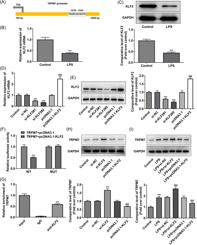
Methods: To establish an in vitro model of S-AKI, RTECs were treated with LPS. The effect of TRPM7 knockdown on cell viability, lactate dehydrogenase (LDH) release, apoptosis, inflammation, and oxidative stress was studied. The binding site between Kruppel-like factor 2 (KLF2) and TRPM7 was predicted using JASPAR. The influence of KLF2 on the regulatory roles of TRPM7 in cells, as well as the effect of their knockdown on the MAPK signaling pathway, was investigated.

Results: TRPM7 was upregulated in LPS-treated cells, and knocking improved cell viability, reduced LDH levels, and minimized apoptosis, inflammation, and oxidative stress. KLF2 was shown to be associated with TRPM7 and its level decreased in LPS-treated cells. KLF2 knockdown increased TRPM7 expression and reversed the effects of TRPM7 knockdown in LPS-treated cells, including suppression of p38 MAPK, ERK1/2, and JNK activation.

Conclusion: Taken together, our results show that TRPM7 is negatively regulated by KLF2 and promotes LPS-induced inflammatory dysfunction by activating the MAPK pathway in RTECs. The theoretical foundation for the prevention and management of S-AKI is laid out in this article.

Keywords: Kruppel-like factor 2; TRPM7; inflammation; renal tubular epithelial cells; sepsis.


Conflict of interest statement

The authors declare no conflicts of interest.

Figures

Figure 1
Figure 1
TRPM7 knockdown increases the viability in lipopolysaccharide (LPS)‐treated cells. (A) The level of TRPM7 in the control and LPS‐treated cells was determined using quantitative real‐time polymerase chain reaction (RT‐qPCR) and (B) western blot analysis. (C) The level of TRPM7 in the transfected cells was detected using RT‐qPCR and (D) western blot analysis. (E) The viability in each group was determined using a Cell Counting Kit‐8 (CCK8) assay. (F) The level of lactate dehydrogenase was measured with an assay kit. ***p < .001 versus control; ## p < .01, ### p < .001 versus LPS + si‐negative control.
Figure 2
Figure 2
TRPM7 knockdown suppresses apoptosis in lipopolysaccharide (LPS)‐treated cells. (A) Cell apoptosis level in each group was evaluated using Terminal deoxynucleotidyl transferase dUTP Nick‐End Labeling assay. (B) The levels of apoptosis‐related proteins were determined using western blot analysis. ***p < .001 versus control; ### p < .001 versus LPS +si‐negative control.
Figure 3
Figure 3
TRPM7 knockdown reduces the inflammation and oxidative stress in lipopolysaccharide (LPS)‐treated cells. (A) The levels of inflammatory factors were determined using an enzyme‐linked immunosorbent assay. (B) The level of cyclooxygenase 2 and inducible nitric oxide synthase were determined using western blot analysis. (C) The oxidative stress level in the cells was measured using assay kits. ***p < .001 versus control; ## p < .01, ### p < .001 versus LPS+si‐negative control.
Figure 4
Figure 4
KLF2 transcription inhibits TRPM7. (A) The binding site between Kruppel‐like factor 2 (KLF2) and TRPM7 promoter was predicted in the JASPAR database. (B) The level of KLF2 in the liposaccharide (LPS)‐treated cells was determined using quantitative real‐time polymerase chain reaction (RT‐qPCR) and (C) western blot analysis. ***p < .001 versus control. (D) The transfection efficacy was verified with RT‐qPCR and (E) western blot analysis. ***p < .001 versus si‐NC; ### p < .001 versus pcDNA3.1. (F) Promoter activity was detected by a luciferase reporter assay. ***p < .001 versus wild‐type (WT)‐TRPM7+pcDNA3.1. (G) The combining capacity between KLF2 and TRPM7 promotors was assessed using a chromatin immunoprecipitation (ChIP) assay. ***p < .001 versus lgG. (H) The level of TRPM7 in the transfected cells without or (I) with LPS treatment was determined using western blot analysis. ***p < .001 versus control or si‐negative control (NC); ### p < .001 versus pcDNA3.1 or LPS + si‐NC; p@@<.01 versus LPS + pcDNA3.1.
Figure 5
Figure 5
KLF2 knockdown reverses the effects of TRPM7 knockdown on cell viability and apoptosis. (A) The viability in each group was determined using a Cell Counting Kit‐8 assay. (B) The level of lactate dehydrogenase release was measured with an assay kit. (C) Cell apoptosis level in each group was evaluated using terminal deoxynucleotidyl transferase dUTP nick‐end labeling assay. (D) The levels of apoptosis‐related proteins were determined using western blot analysis. ***p < .001 versus control; ## p < .01, ### p < .001 versus liposaccharide (LPS); @ p < .05, p@@@<.001 versus LPS+si‐TRPM7+si‐negative control.
Figure 6
Figure 6
KLF2 knockdown reverses the effects of TRPM7 knockdown on inflammation, oxidative stress, and p38 MAPK signaling. (A) The levels of inflammatory factors were determined using an enzyme‐linked immunosorbent assay (ELISA). (B) The level of cyclooxygenase 2 and inducible nitric oxide synthase (iNOS) were determined using western blot analysis. (C) The oxidative stress level in the cells was measured using assay kits. (D) The abundance of p38 MAPK, ERK1/2, JNK, and their phosphorylated forms in these groups of cells were determined using western blot analysis. ***p < .001 versus control; ### p < .001 versus liposaccharide; @ p < .05, p@@<.01, p@@@<.001 versus LPS + si‐TRPM7+si‐negative control.

References

    1. Salomão R, Ferreira BL, Salomão MC, Santos SS, Azevedo LCP, Brunialti MKC. Sepsis: evolving concepts and challenges. Braz. J. Med. Biol. Res. 2019;52(4):e8595. 10.1590/1414-431x20198595 - DOI - PMC - PubMed
    1. Thompson K, Venkatesh B, Finfer S. Sepsis and septic shock: current approaches to management. Intern Med J. 2019;49(2):160‐170. 10.1111/imj.14199 - DOI - PubMed
    1. Fleischmann C, Scherag A, Adhikari NK, et al. Assessment of global incidence and mortality of hospital‐treated sepsis. Current estimates and limitations. Am J Respir Crit Care Med. 2016;193(3):259‐272. 10.1164/rccm.201504-0781OC - DOI - PubMed
    1. Perner A, Gordon AC, De Backer D, et al. Sepsis: frontiers in diagnosis, resuscitation and antibiotic therapy. Intensive Care Med. 2016;42(12):1958‐1969. 10.1007/s00134-016-4577-z - DOI - PubMed
    1. Mirouse A, Vigneron C, Llitjos JF, et al. Sepsis and cancer: an interplay of friends and foes. Am J Respir Crit Care Med. 2020;202(12):1625‐1635. 10.1164/rccm.202004-1116TR - DOI - PubMed

Publication types