. 2022 Jul;10(7):e667.
doi: 10.1002/iid3.667.

Antiviral activity of triptolide on herpes simplex virus in vitro

Affiliations

Antiviral activity of triptolide on herpes simplex virus in vitro

Nasrin Aliabadi et al. Immun Inflamm Dis. 2022 Jul.

Abstract

Background: Herpes simplex virus-type 1 (HSV-1) can cause diseases, especially amongst neonates and immunocompromised hosts. Hence, developing a novel anti-HSV-1 drug with low-level toxicity is vital. Triptolide (TP), a diterpenoid triepoxide is a natural product with range of bioactivity qualities.

Methods: In this study, viral infection was assessed in different phases of the HSV-1 replication cycle on A549 cells, using various assays, such as adsorption inhibition assay, penetration inhibition assay, time-of-addition assay, and quantitative polymerase chain reaction (qPCR).

Results: The results indicate that TP can effectively inhibit HSV-1 infection in the lowest range of concentration. TP exhibited significant inhibitory effect on HSV-1 plaque formation, with 50% effective concentration (EC50) of 0.05 µM. Furthermore, the time-of-addition assay suggests that TP has viral inhibitory effects when it was added less than 8 h postinfection (h.p.i.). This result is further confirmed by decline in the expression viral immediate-early genes (ICP4, ICP22, and ICP27) in 6 h.p.i in the TP-treated group compared to the control group, evaluated by real-time qPCR. The Western blotting result was also consistent with the previous findings, which confirms that TP can positively affect ICP4 during HSV-1 infection.

Conclusions: The TP also showed antiviral activity against HSV-1. This dose-dependent activity is an indication of a particular cellular component, rather than cytotoxicity that has mediated its function. Finally, the result suggest a new approach for an effective treatment option of the HSV-1 infections.

Keywords: HSV-1; ICP4; antiviral; natural product; triptolide.


Conflict of interest statement

The authors declare no conflict of interest.

Figures

Figure 1
Figure 1
The mechanism of the triptolide (TP)‐mediated inhibition of herpes simplex virus‐type 1 (HSV‐1). Direct virucidal effect test. HSV‐1 was mixed with TP (6 × EC50% µM), acyclovir (ACV) (6 ×  EC50% µM), or DMSO (0.15%%) (consisting of DMSO, virus, and cells) and was then incubated at room temperature (25°C) for 1 h. The residual infectious virus titers were demonstrated by the plaque assay. The data were obtained from three independent experiments. Error bars indicate standard deviations from the three independent experiments.
Figure 2
Figure 2
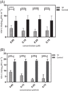
(A) The effect of triptolide (TP) on virus adsorption and (B) penetration. This procedure was performed as described in Section 2. The data represent the results of three independent experiments that were performed repeatedly. Statistical significance between the compound‐treated and DMSO‐treated groups was determined by the Student's t‐test: ns; ***p < .001; ****p < .0001.
Figure 3
Figure 3
The time‐of‐addition assay. A549 cells were infected with herpes simplex virus‐type 1 (HSV‐1) at the MOI of 0.1. Then, triptolide (TP) (3 × EC50% µM), ACV (0.01 μg/ml), and 0.15% DMSO were added to the cells at the indicated time points both before and after infection. The infected cells were lysed by performing three cycles of freezing and thawing. The residual infectivity of the treated viruses in different time courses was further investigated by titration on the Vero cells. The values were obtained by the virus yield titration of cell lysates and represented the mean of an independent experiment in triplicate (±SE). *** indicates significant differences between the tested sample and DMSO (p < .0001). ANOVA/Dunnett's tests were carried out, as appropriate. ACV, acyclovir.
Figure 4
Figure 4
Quantitative polymerase chain reaction was performed to assess the number of herpes simplex virus‐type 1 (HSV‐1) genome copies after treatment with triptolide (TP) (a) in the presence or absence of these compounds compared to the DMSO control (consisting of the virus, 0.15% DMSO, and cells). The data have been presented as mean and standard deviation. Significant differences were found between the TP and DMSO control groups (consisting of the virus, 0.15% DMSO, and cells) regarding the HSV‐1 DNA level up to 8 h.p.i. *** indicates significant differences between the tested sample and the virus control (p < .0001). ANOVA/Dunnett's tests were carried out, as appropriate.
Figure 5
Figure 5
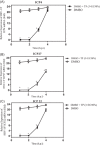
The effect of triptolide (TP) (3 × EC50% µM) on the expression of herpes simplex virus‐type 1 (HSV‐1) (immediate‐early genes [IE]) (A)–(C). A549 cells were infected with HSV‐1 at the MOI of 0.1 and were then treated with and without 3 × EC50% µM of the TP compound. RNA was extracted at specified intervals and real‐time quantitative polymerase chain reaction was done using mRNA‐specific primers. Gene transcription level was defined as relative based on the mRNA fold change (2Ct) at each time point. It was initially normalized by GAPDH and was then compared to the DMSO group (consisting of the virus, 0.15% DMSO, and cells) at 4 h.p.i. ANOVA/Dunnett's tests were carried out, as appropriate.
Figure 6
Figure 6
For western blot analysis of ICP4, the cells grown in a six‐well plate were infected with HSV‐1 at the MOI of 0.01 and were treated with or without 3 × EC50 μM TP. Then, the cells were harvested. There was a noticeable difference between the 3 × EC50 μM TP and 0.15% DMSO groups at 4 and 6 h.p.i. Molecular weight standards in kiloDaltons are indicated to the left of each panel.

References

    1. Fields BN. Fields' virology. Vol 1. Lippincott Williams & Wilkins; 2007.
    1. Harkness JM, Kader M, DeLuca NA. Transcription of the herpes simplex virus 1 genome during productive and quiescent infection of neuronal and nonneuronal cells. J Virol. 2014;88(12):6847‐6861. - PMC - PubMed
    1. Aliabadi N, Jamalidoust M, Asaei S, Namayandeh M, Ziyaeyan M. Diagnosing of herpes simplex virus infections in suspected patients using real‐time PCR. Jundishapur J Microbiol. 2015;8(2):e16727. - PMC - PubMed
    1. Smith JS, Robinson NJ. Age‐specific prevalence of infection with herpes simplex virus types 2 and 1: a global review. J Infect Dis. 2002;186(suppl 1):S3‐S28. - PubMed
    1. Kolokotronis A, Doumas S. Herpes simplex virus infection, with particular reference to the progression and complications of primary herpetic gingivostomatitis. Clin Microbiol Infect. 2006;12(3):202‐211. - PubMed

Publication types