. 2022 Jun 1;57(6):251-259.
doi: 10.1097/SHK.0000000000001934.

Fecal Microbiota Transfer Attenuates Gut Dysbiosis and Functional Deficits After Traumatic Brain Injury

Affiliations

Fecal Microbiota Transfer Attenuates Gut Dysbiosis and Functional Deficits After Traumatic Brain Injury

Booker T Davis 4th et al. Shock. .

Abstract

Background: Traumatic brain injury (TBI) is an underrecognized public health threat. Survivors of TBI often suffer long-term neurocognitive deficits leading to the progressive onset of neurodegenerative disease. Recent data suggests that the gut-brain axis is complicit in this process. However, no study has specifically addressed whether fecal microbiota transfer (FMT) attenuates neurologic deficits after TBI.

Hypothesis: We hypothesized that fecal microbiota transfer would attenuate neurocognitive, anatomic, and pathologic deficits after TBI.

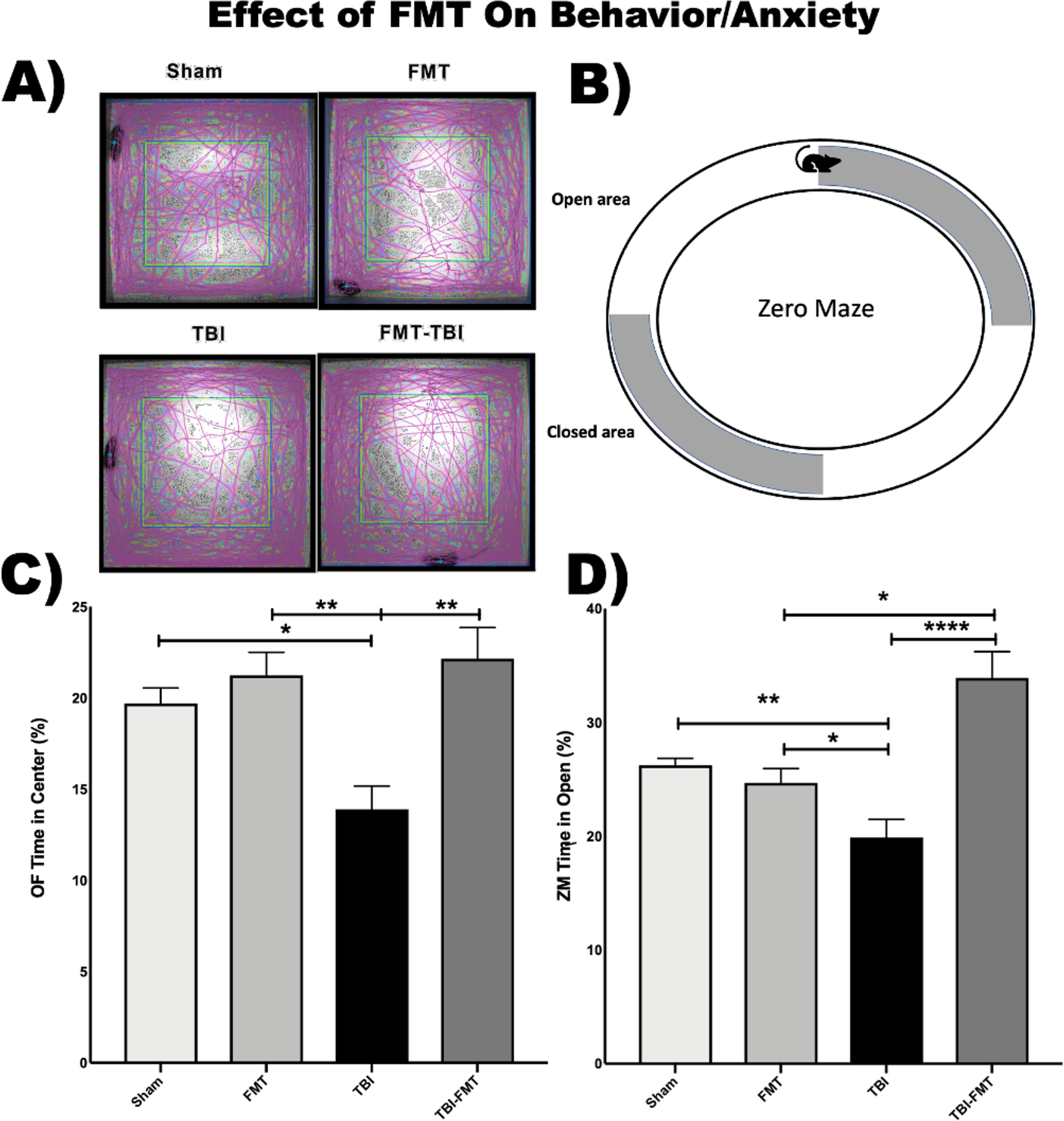
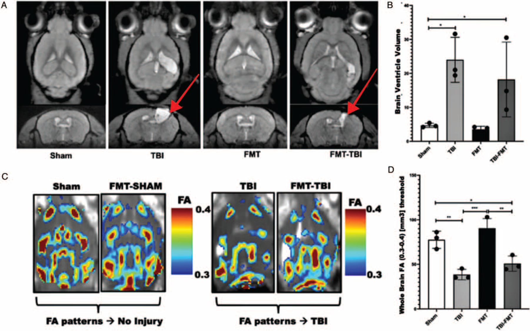
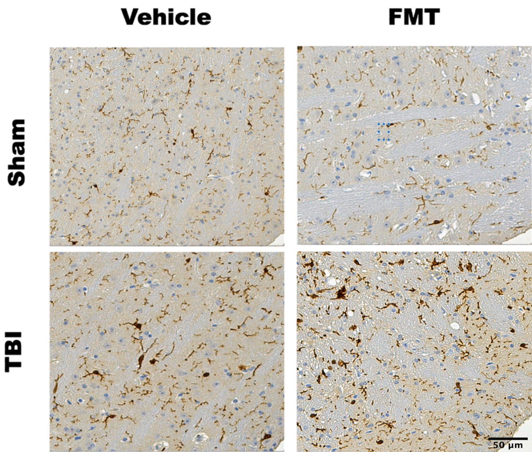
Methods: C57Bl/6 mice were subjected to severe TBI (n = 20) or sham-injury (n = 20) via an open-head controlled cortical impact. Post-injury, this cohort of mice underwent weekly oral gavage with a slurry of healthy mouse stool or vehicle alone beginning 1 h post-TBI followed by behavioral testing and neuropathologic analysis. 16S ribosomal RNA sequencing of fecal samples was performed to characterize gut microbial community structure pre- and post-injury. Zero maze and open field testing were used to evaluate post-traumatic anxiety, exploratory behavior, and generalized activity. 3D, contrast enhanced, magnetic resonance imaging was used to determine differences in cortical volume loss and white matter connectivity. Prior to euthanasia, brains were harvested for neuropathologic analysis.

Results: Fecal microbiome analysis revealed a large variance between TBI, and sham animals treated with vehicle, while FMT treated TBI mice had restoration of gut dysbiosis back to levels of control mice. Neurocognitive testing demonstrated a rescue of normal anxiety-like and exploratory behavior in TBI mice treated with FMT. FMT treated TBI mice spent a greater percentage of time (22%, P = 0.0001) in the center regions of the Open Field as compared to vehicle treated TBI mice (13%). Vehicle-treated TBI animals also spent less time (19%) in the open areas of zero maze than FMT treated TBI mice (30%, P = 0.0001). Comparing in TBI mice treated with FMT, MRI demonstrated a marked attenuation in ventriculomegaly (P < 0.002) and a significant change in fractional anisotropy (i.e., loss of white matter connectivity) (P < 0.0001). Histologic analysis of brain sections revealed a FMT- injury dependent interaction in the microglia/macrophage-specific ionized calcium-binding protein, Iba1 (P = 0.002).

Conclusion: These data suggest that restoring a pre-injury gut microbial community structure may be a promising therapeutic intervention after TBI.


Conflict of interest statement

The authors report no conflict(s) of interest.

Figures

Fig. 1.
Fig. 1.. Transfer of stool from healthy donor mice reverses the loss of species richness and species evenness after TBI in mice.
Fecal microbiome biodiversity was measured and is represented as box plots of species richness (A) and species evenness (B). Elements of alpha-diversity, measured by Simpson Index, which relates operational taxonomic unit (OTU) richness and evenness, are plotted with each box representing groups containing 5 mice (N = 5). TBI results in a marked decrease in the number of species present and their relative abundance. Post-TBI treatment with FMT abrogates TBI-induced decreases in richness and evenness. ((P ≤ 0.05(*), 0.001(**), and 0.0001(***)).
Fig. 2.
Fig. 2.. Transfer of stool from health donor mice preserves the ratio between regional and local species diversity after TBI.
These graphs depict the relative distinctness and similarities of all groups. A principal coordinates analysis (PCoA) diagram (A) shows groups overall similarity of using relative placement along axis PC1 and PC2. Dots in the PCoA plot represent an individual sample. Colors indicate group (Red = Sham vehicle, Blue = TBI vehicle, Green = Sham-FMT, and Purple = TBI-FMT). In the chart, sham and TBI groups are separated by the most distance representing the greatest level of between-groups-variability. Despite distance, these two groups show similar clustering representing the least amount of within-groups-variability of microbial phenotypes. The Atchison graph depicts the internal group variability (mean) using nodes (polygons) of large or small sizes to represent the level of internal group diversity. Distance of nodes from a central point (centroid) reveals group similarity of group means. This Atchison plot reveals that sham-FMT samples show the greatest within group diversity while the small TBI polygon depicts the least. Similar sized Sham and FMT-TBI nodes have similar levels of internal group diversity.
Fig. 3.
Fig. 3.. Murine fecal microbiome bacterial species post TBI and FMT treatment.
Heatmap plots were generated to represent the species-level differences in the microbial community of all groups at 59 days post TBI. Experimental groups are highlighted along the X-axis. Measured species are along the Y-axis. Color shaded lines represent the abundance of each species found with the most abundant species within a group in red and least abundant species shown with a yellow line. The graph reveals the presence of group dependent microbial networks due to treatment and injury.
Fig. 4.
Fig. 4.. Fecal microbiome transfer alters anxiety and exploratory behavior in TBI mice.
(A) Open Field (OF) was used to assess anxiety and exploratory behavior. The purple lines in this panel represent the path taken by mice in the five-minute testing period. (B) Elevated Zero Maze (ZM) was used to assess for post-injury anxiety. More time spent in the open areas indicates a higher inclination towards exploration and a lower level of less anxiety. (C) OF testing demonstrated preserved exploratory behavior in TBI mice post injury treatment as demonstrated by the increased percentage of time spent in the center of the open field compared to untreated injured animals (22% vs 19%: N = 5/group). (D) ZM demonstrated preservation of normal anxiety-like behavior in FMT-treated TBI mice as compared to vehicle-treated TBI mice with a significant increase in time spent in the open area (39% vs 19%; P = 0.0001; N = 5/group).
Fig. 5.
Fig. 5.. FMT attenuates cortical volume loss and preserves white matter connectivity after TBI.
(A) Representative longitudinal and transverse MRI scans of animals (60DPI) with injury sites indicated with red arrows (N = 3). (B) FMT attenuates TBI-induced ventriculomegaly (red arrows) which is a well-described surrogate for cortical volume loss (P < 0.002). (C) Images ktrans level (measure of capillary permeability) were extracted from contrast-enhanced 3D-MRI. Fractional anisotropy (connectivity) maps were then extracted from the MRI images. (D) TBI induced a decrease in fractional anisotropy (white matter connectivity) compared to sham injury (P < 0.0001). FMT treatment attenuated this loss of connectively as compared to vehicle treated TBI mice (P = 0.04).
Fig. 6.
Fig. 6.. Ionized calcium binding adaptor molecule 1 (IBA1) stained sections of ipsilateral mouse cortex.
IBA1 stained tissue from the ipsilateral cortex at the impact site 90 days after localized CCI brain injury. Ten micrometers of samples from six equalized locations within the ipsilateral cortex of TBI and Sham-TBI brains +/− FMT treatment at the 90DPI timepoint. Sham injured mice show no appreciable increase in IBA1 staining whether treated with FMT or vehicle. TBI resulted in a marked increase in IBA1 staining as compared to sham injury. Post injury treatment with FMT resulted in a further increase in IBA1 staining as compared to vehicle treatment TBI mice (P = 0.002).

References

    1. Nittayasoot N, Peterson AB, Thammawijaya P, Parker EM, Sathawornwiwat A, Boonthanapat N, Chantian T, Voradetwitaya L, Jiraphongsa C, Sagarasaeranee O, et al.: Evaluation of a hospital-based injury surveillance system for monitoring road traffic deaths in Phuket, Thailand. Traffic Inj Prev 20(4):365–371, 2019. - PMC - PubMed
    1. DeKosky ST, Asken BM: Injury cascades in TBI-related neurodegeneration. Brain Inj 31(9):1177 –1182, 2017. - PMC - PubMed
    1. Faul M, Coronado V. Chapter 1: Epidemiology of traumatic brain injury In: Grafman J, Salazar AM, eds. Vol. 127. Elsevier, 2015, 3–13. - PubMed
    1. Makinde HM, Cuda CM, Just TB, Perlman HR, Schwulst SJ: Nonclassical monocytes mediate secondary injury, neurocognitive outcome, and neutrophil infiltration after traumatic brain injury. J Immunol 199(10):3583 –3591, 2017. - PMC - PubMed
    1. McAllister TW: Neurobiological consequences of traumatic brain injury. Dialogues Clin Neurosci 13(3):287–300, 2011. - PMC - PubMed

Publication types