Review
. 2022 Oct 6:38:1-23.
doi: 10.1146/annurev-cellbio-120420-114559. Epub 2022 Jun 27.

How Microtubules Build the Spindle Branch by Branch

Affiliations
Review

How Microtubules Build the Spindle Branch by Branch

Sophie M Travis et al. Annu Rev Cell Dev Biol. .

Abstract

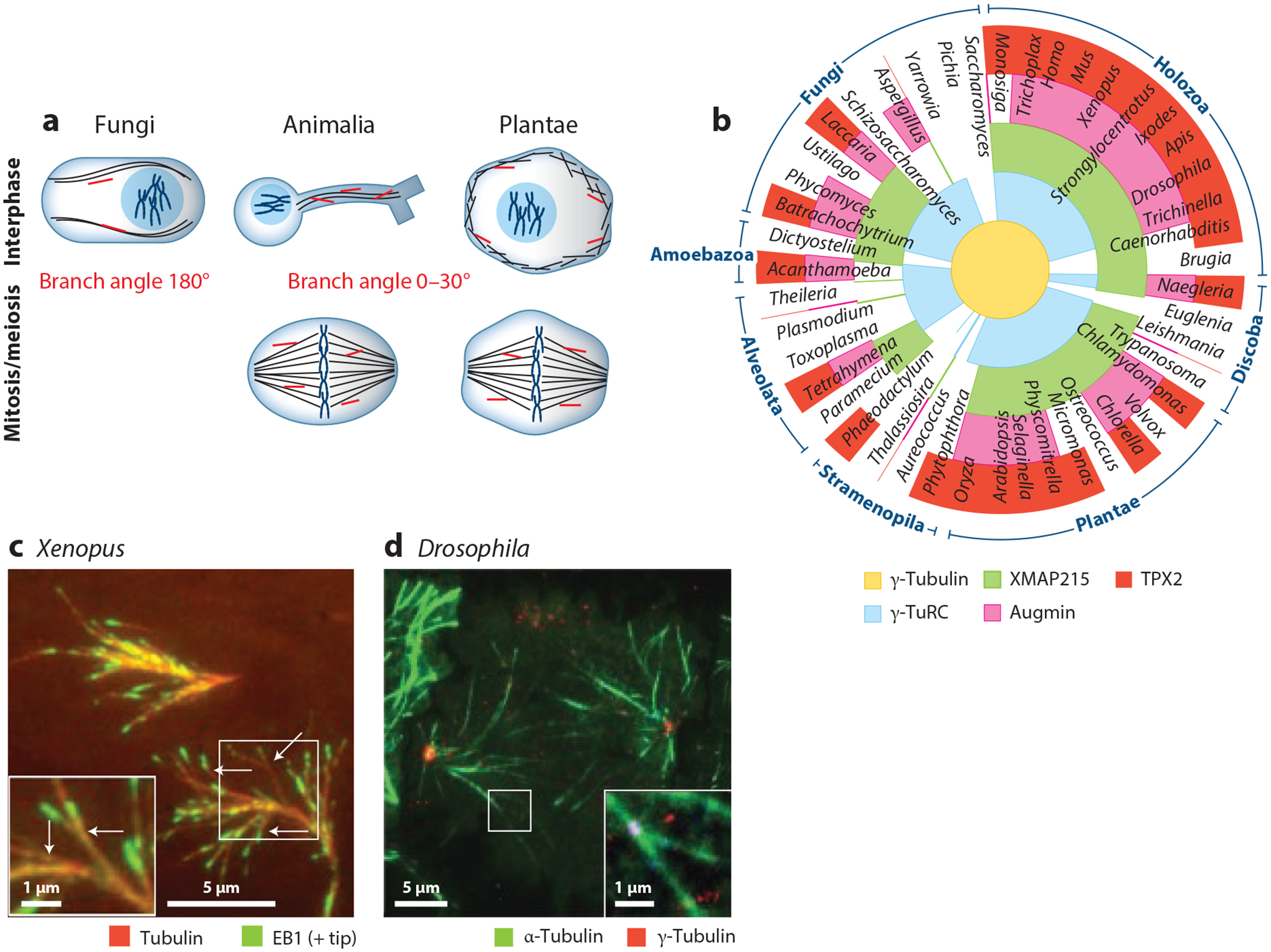
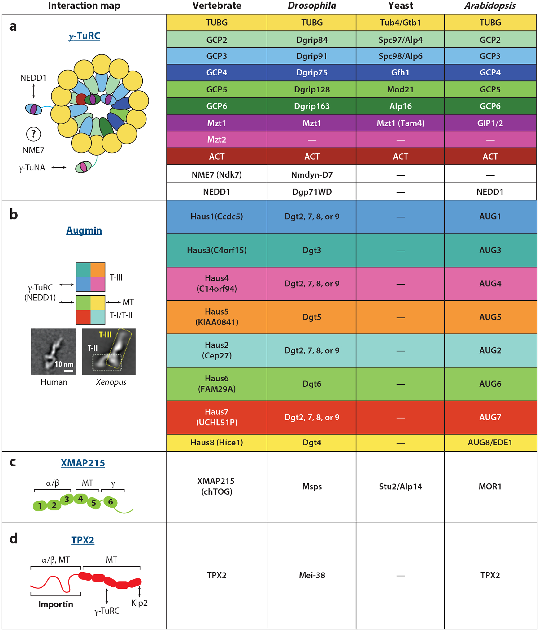
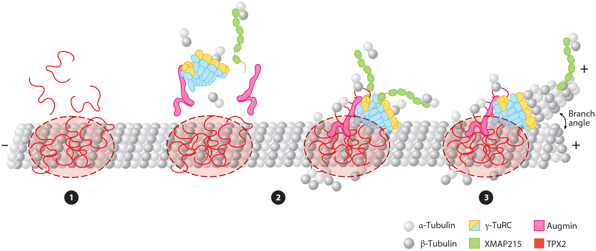
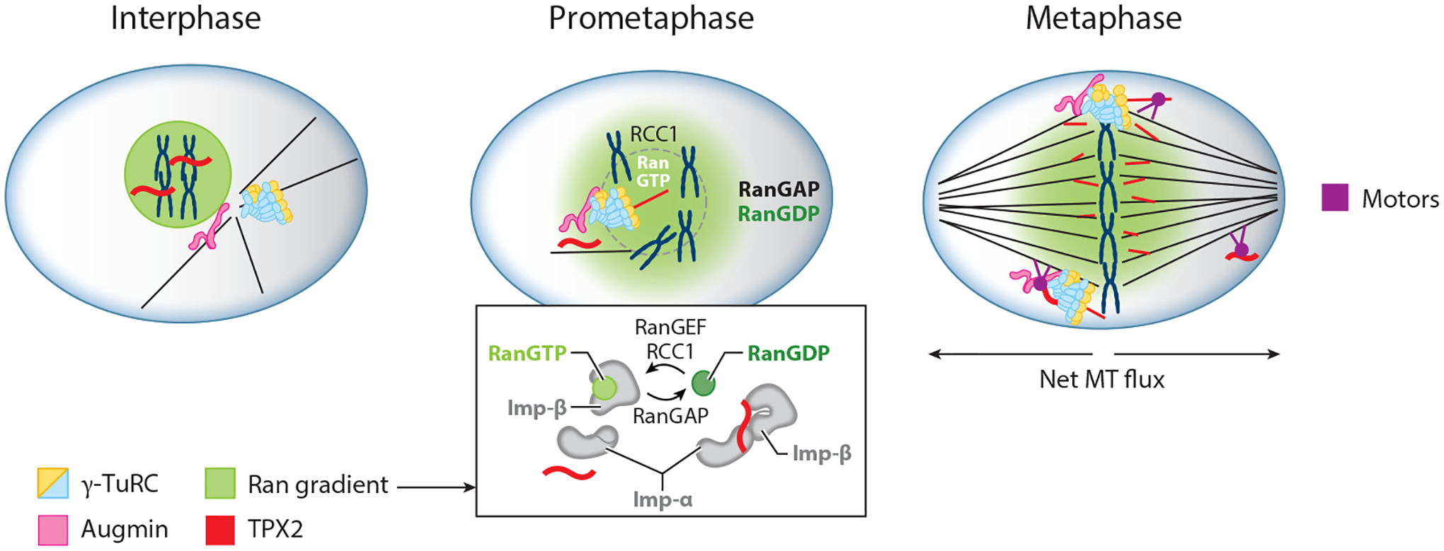
The microtubule (MT) cytoskeleton provides the architecture that governs intracellular organization and the regulated motion of macromolecules through the crowded cytoplasm. The key to establishing a functioning cytoskeletal architecture is regulating when and where new MTs are nucleated. Within the spindle, the vast majority of MTs are generated through a pathway known as branching MT nucleation, which exponentially amplifies MT number in a polar manner. Whereas other MT nucleation pathways generally require a complex organelle such as the centrosome or Golgi apparatus to localize nucleation factors, the branching site is based solely on a simple, preformed MT, making it an ideal system to study MT nucleation. In this review, we address recent developments in characterizing branching factors, the branching reaction, and its regulation, as well as branching MT nucleation in systems beyond the spindle and within human disease.

Keywords: TPX2; augmin; branching microtubule nucleation; meiosis; mitosis; γ-TuRC; γ-tubulin ring complex.


Figures

Figure 1
Figure 1
Branching microtubule (MT) nucleation is conserved across most eukaryotes and cell types. (a) Branching MT nucleation has been studied in both interphase and mitosis/meiosis across fungi, animals, and plants. MT branches are displayed in orange, and the ranges of branch angles relative to the parent MT are annotated at the center, where at 0° the two MTs are parallel and at 180° they are antiparallel. (b) Diverse eukaryotic species are represented as spokes in a circular eukaryotic phylogeny, in which phylum groupings are annotated on the exterior of the circle. The radial sections of each spoke are colored based on the presence or absence of nucleation and branching factors: from the center, γ-tubulin (yellow), γ-tubulin ring complex (γ-TuRC) subunit γ-tubulin complex protein 4 (GCP4) (blue), Xenopus MT-associated protein of 215 kDa (XMAP215) with a tumor overexpressed gene 6 (TOG6) domain to enable binding to γ-TuRC (green), augmin subunit Haus6 (pink), and targeting protein for Xklp2 (TPX2) (red). (c) Ex vivo visualization of branching MT nucleation in meiotic Xenopus laevis egg extract system. MTs are labeled in red via Alexa568 tubulin and growing plus ends via green fluorescent protein (GFP)-end-binding protein 1 (EB1). Arrows indicate branch sites. Panel c reproduced from Petry et al. (2013). (d) Visualization of MT branching in mitotic Drosophila melanogaster S2 cells during anaphase. Microtubules are labeled in green via GFP-α-tubulin and branch sites via γ-tubulin-red fluorescent protein (RFP). Panel d reproduced with permission from Verma & Maresca (2019).
Figure 2
Figure 2
Orthologous branching factors across model organisms. (a) The nucleator γ-TuRC consists of a spiral of 14 γ-tubulins scaffolded by GCPs. Mzt/GCP complexes occupy diverse positions throughout the ring, binding the accessory subunit NEDD1 and the localizer/activator γ-TuNA. The cartoon of γ-TuRC is based on data from Consolati et al. (2020), Liu et al. (2020), and Wieczorek et al. (2020a,b). (b) The augmin complex is an “h” shaped hetero-octamer separable into the MT-binding tetramer I/II (T-I/II) and γ-TuRC-binding tetramer III (T-III). Augmin is lost in yeast, and low sequence homology prevents four of the eight Drosophila augmin subunits (Dgt2, Dgt7, Dgt8, and Dgt9) from being matched to their vertebrate and plant counterparts. The 2D classification of human augmin was reproduced with permission from Hsia et al. (2014); the 2D classification of Xenopus augmin was reproduced from Song et al. (2018). (c) The microtubule polymerase and nucleator XMAP215 consists of multiple tubulin-binding TOG domains, where TOG1–3 bind soluble tubulin, TOG4–5 bind polymerized tubulin, and TOG6 binds γ-tubulin. (d) TPX2, a large, poorly-ordered protein, binds tubulin in various forms, as well as itself, γ-TuRC, and importin regulatory proteins. The cartoon of TPX2 is based on data from Alfaro-Aco et al. (2017). Abbreviations: γ-TuNA, γ-tubulin nucleator activator; γ-TuRC, γ-tubulin ring complex; ACT, actin; Alp, altered polarity; AUG, augmin; CCDC, coiled-coil domain containing protein; Cep, centrosomal protein; chTOG, colonic and hepatic tumor overexpressed gene; Dgp, Drosophila γ-ring protein; Dgt, dim γ-tubulin; Dgrip, Drosophila γ-ring protein; EDE, endosperm defective; FAM, family with sequence similarity; GCP, γ-tubulin complex protein; Gfh, GCP4 homolog; GIP, GCP3-interacting protein; Haus, homologous to augmin subunit; Hice, Hec1-interacting and centrosome-associated; KIAA, Kazusa cDNA sequencing project gene; Klp, kinesin-like protein; Mei, meiotic mutant; Mod, morphology defective; MOR, microtubule organization; Msps, minispindles; MT, microtubule; Mzt, mitotic spindle organizing proteins associated with a ring of γ-tubulin; NEDD, neural precursor cell expressed, developmentally-downregulated; NME, non-metastatic cells; Spc, spindle pole component; Stu, suppressor of tubulin; Tam, transcript altered in meiosis; TPX, targeting protein for Xenopus kinesin-like protein; TUB, tubulin; UCHL5, ubiquitin carboxy-terminus hydrolase isozyme L5; XMAP215, Xenopus MT-associated protein of 215 kDa.
Figure 3
Figure 3
The temporal order of branching microtubule (MT) nucleation. ❶ TPX2 condenses into a liquid-like phase on the MT. ❷ Soluble tubulin and branching factors are recruited to the targeting protein for Xklp2 (TPX2) droplet. Augmin and Xenopus MT-associated protein of 215 kDa (XMAP215) may be recruited individually or in complex with γ-tubulin ring complex (γ-TuRC), and additional soluble tubulin may be recruited by XMAP215 and γ-TuRC. ❸ γ-TuRC with XMAP215 nucleates a new MT at an acute angle to the parent MT. After nucleation of the daughter MT, some molecules of XMAP215 track the plus tip of the growing MT.
Figure 4
Figure 4
Branching MT nucleation is regulated within spindle assembly. In interphase, branching is inactive due to sequestration of TPX2 (red) within the nucleus by RanGTP. After nuclear envelope breakdown in prometaphase, RanGTP and active TPX2 are released to initiate branching near chromosomes due to the presence of RanGEF RCC1. The inset shows how RanGTP (green), acting through Imp-α and Imp-β, releases TPX2, and RanGDP (green-gray) causes TPX2 to be sequestered. During metaphase, the interaction of motors (purple) with TPX2 and MT branches causes both TPX2, new MTs, and perhaps other branching factors to relocate throughout the spindle. Abbreviations: γ-TuRC, γ-tubulin ring complex; Imp-α/β, importin α/β; MT, microtubule; RanGAP, Ran GTPase-activating protein; RanGEF, Ran guanine nucleotide exchange factor; RCC1, regulator of chromosome condensation 1; TPX2, targeting protein for Xklp2.

References

    1. Alfaro-Aco R, Thawani A, Petry S. 2017. Structural analysis of the role of TPX2 in branching microtubule nucleation. J. Cell Biol 216:983–97 - PMC - PubMed
    1. Alfaro-Aco R, Thawani A, Petry S. 2020. Biochemical reconstitution of branching microtubule nucleation. eLife 9:e49797. - PMC - PubMed
    1. Alvarado-Kristensson M 2018. γ-tubulin as a signal-transducing molecule and meshwork with therapeutic potential. Signal Transduct. Target. Ther 3:24. - PMC - PubMed
    1. Boissan M, Schlattner U, Lacombe ML. 2018. The NDPK/NME superfamily: state of the art. Lab. Investig 98:164–74 - PubMed
    1. Borek WE, Groocock LM, Samejima I, Zou J, de Lima Alves F, et al. 2015. Mto2 multisite phosphorylation inactivates non-spindle microtubule nucleation complexes during mitosis. Nat. Commun 6:7929. - PMC - PubMed

Publication types

MeSH terms

LinkOut - more resources