. 2022 Jun 27;12(1):10877.
doi: 10.1038/s41598-022-14860-0.

Evidence for mitochondrial Lonp1 expression in the nucleus

Affiliations

Evidence for mitochondrial Lonp1 expression in the nucleus

Lara Gibellini et al. Sci Rep. .

Abstract

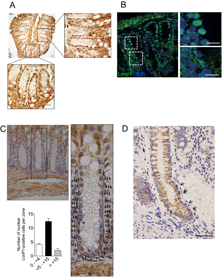
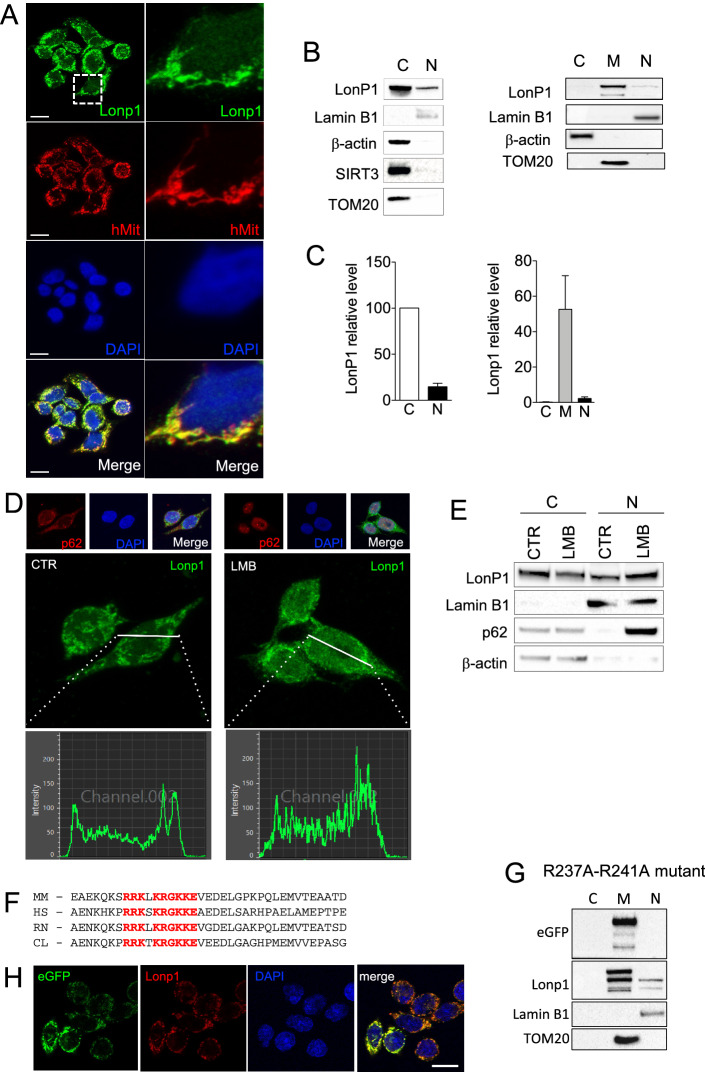
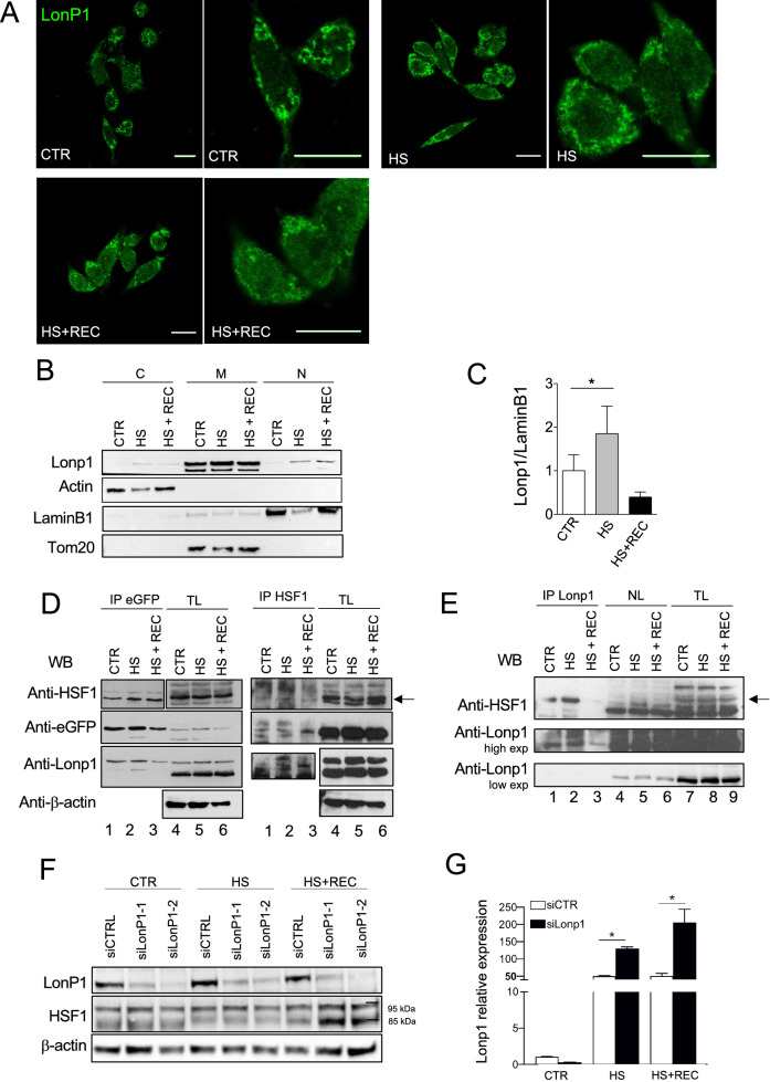
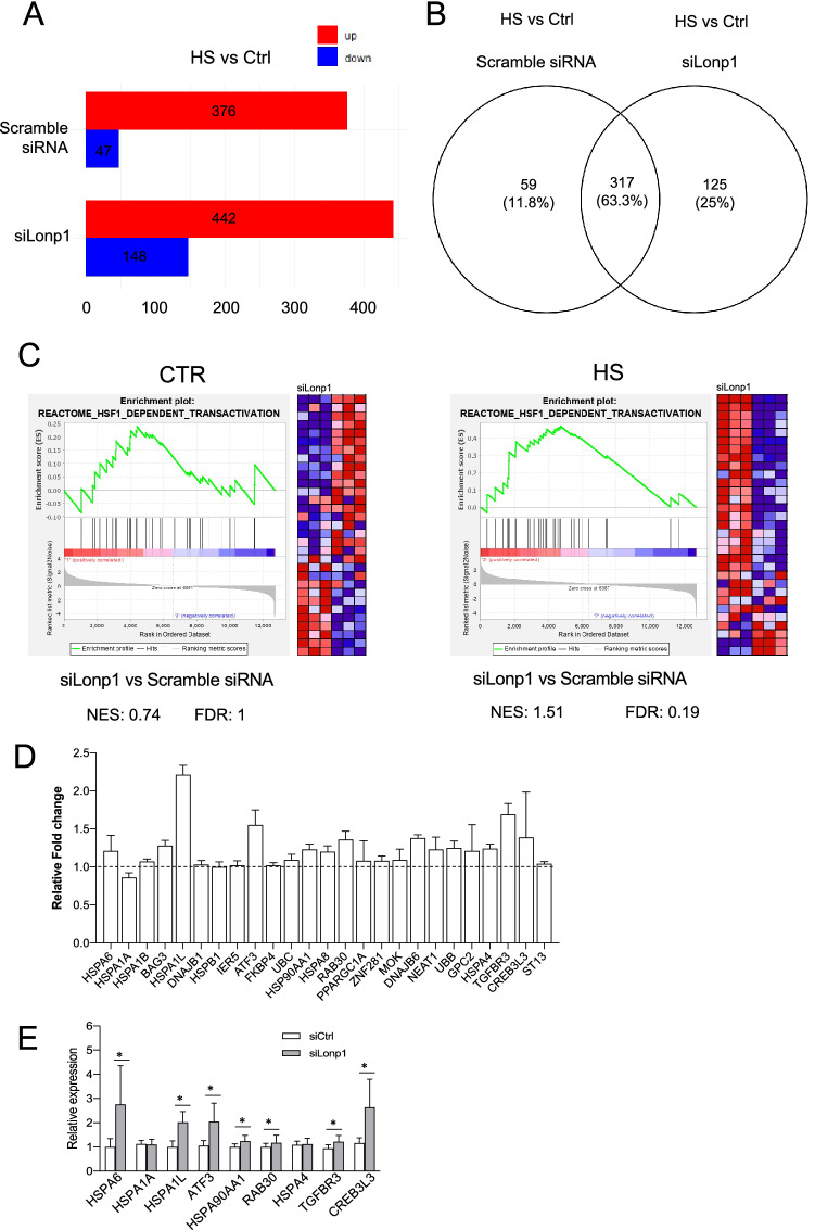
The coordinated communication between the mitochondria and nucleus is essential for cellular activities. Nonetheless, the pathways involved in this crosstalk are scarcely understood. The protease Lonp1 was previously believed to be exclusively located in the mitochondria, with an important role in mitochondrial morphology, mtDNA maintenance, and cellular metabolism, in both normal and neoplastic cells. However, we recently detected Lonp1 in the nuclear, where as much as 22% of all cellular Lonp1 can be found. Nuclear localization is detectable under all conditions, but the amount is dependent on a response to heat shock (HS). Lonp1 in the nucleus interacts with heat shock factor 1 (HSF1) and modulates the HS response. These findings reveal a novel extramitochondrial function for Lonp1 in response to stress.


Conflict of interest statement

The authors declare no competing interests.

Figures

Figure 1
Figure 1
Lonp1 localizes in the nuclei of mouse colon tissue and human colon tissue. (A) Representative images of Lonp1 IHC on murine colon tissue, showing Lonp1 nuclear localization. (B) Representative images of Lonp1 IF on murine colon tissue, showing Lonp1 nuclear localization. Nuclei were counterstained with DAPI. (C) Representative Ki-67 IHC staining in intestinal crypt. Cells of the crypt were sectioned into three zones: the lower (0 to + 4), upper (+ 5 to + 15), and non (> + 15)-crypt zones. The histogram represents the percentage of cells with nuclear Lonp1 per crypt zone (n = 10 crypts per group). Data represent the mean ± SD. (D) Representative images of Lonp1 IHC on human colon tissue, showing Lonp1 nuclear localization (arrows). Bar: 200 µm.
Figure 2
Figure 2
A fraction of Lonp1 localizes in the nuclei of human cells. (A) Representative confocal microscopy images of SW620 cells after immunostaining with anti-Lonp1 and anti-human mitochondria (hMit) Abs. Nuclei were counterstained with DAPI. Bars: 10 mm. (B) Left panel: representative immunoblot of cytosolic (C) and nuclear (N) fractions obtained from SW620 cells. Lamin B1 is the nuclear fraction loading control, whereas β-actin is the cytosolic fraction loading control. Immunoblots of Sirtuin-3 (SIRT3) and TOM20 are also reported to indicate that mitochondrial contamination is not present. Blots were cut prior to hybridisation with antibodies during blotting. Right panel: representative immunoblot of cytosolic (C), mitochondrial (M), nuclear (N) fractions obtained from SW620 cells. β-actin is the cytosolic fraction loading control, TOM20 is the mitochondrial fraction loading control and lamin B1 is the nuclear fraction loading control. (C) Left histogram: quantification of the ratio of nuclear and cytosolic Lonp1 (n = 3 independent experiments). Right histogram: quantification of the ratio of cytosolic, mitochondrial and nuclear Lonp1 (n = 3 independent experiments). (D) Representative confocal microscopy images of SW620 cells after immunostaining with anti-Lonp1 antibody. Cells were treated or not with leptomycin B (LMB) to inhibit nuclear export of proteins. Staining of p62 was reported as positive control for LBM treatment. Nuclei were counterstained with DAPI. (E) Representative immunoblot of cytosolic (cytosol) and nuclear (N) fractions obtained from SW620 cells, treated or not with LBM. Lamin B1 is the nuclear fraction loading control, whereas β-actin is the cytosolic fraction loading control. Immunoblot for p62 is also reported as positive control for LMB treatment. Blots were cut prior to hybridisation with antibodies during blotting. (F) Alignment of Lonp1 protein sequences (amino acids 218 to 256) from the indicated mammalian species. HS, Homo sapiens; CL, Canis lupus familiaris; MM, Mus musculus; RN, Rattus norvegicus. The nuclear targeting sequence is highlighted in red. (G) Representative immunoblot of cytosolic (C), mitochondrial (M), nuclear (N) fractions obtained from SW620 cells expressing the R237A-R241A mutant of Lonp1. β-actin is the cytosolic fraction loading control, TOM20 is the mitochondrial fraction loading control and lamin B1 is the nuclear fraction loading control. Blots were cut prior to hybridisation with antibodies during blotting. (H) Representative confocal microscopy images of SW620 cells expressing the R237A-R241A mutant of eGFP-Lonp1 after immunostaining with anti-Lonp1 antibody. Nuclei were counterstained with DAPI. Bar: 10 mm.
Figure 3
Figure 3
Lonp1 relocates in the nucleus in response to heat shock. (A) Representative confocal microscopy images of SW620 cells after immunostaining with anti-Lonp1 antibody. Cells were kept at 37 °C (CTRL), heat-shocked (HS) at 42 °C and then left to recover (HS + REC). Bars: 10 mm. (B) Representative immunoblot showing Lonp1 expression in cytosolic, mitochondrial and nuclear fractions from SW620 cells maintained at 37 °C (CTR), at 42 °C for 3 h to induce heat shock (HS) and kept at 42 °C for 3 h and then left for 1 h at 37 °C to recover (HS + REC). β-actin is the cytosolic fraction loading control, TOM20 is the mitochondrial fraction loading control and lamin B1 is the nuclear fraction loading control. Blots were cut prior to hybridisation with antibodies during blotting. (C) Histogram representing the relative levels of Lonp1 in nuclear fractions, obtained from three independent experiments. Data are shown as mean SD. *p < 0.05. (D) Representative immunoprecipitation experiment showing the interaction between Lonp1-eGFP and HSF1 in SW620 cells after heat-shock (HS) and then left 3 h at 37 °C to recover (HS + REC). Left panels: lysates were immunoprecipitated with anti-eGFP and immunoblotted with anti-HSF1. Western blot on total lysate (TL) is also shown. Blots were cut prior to hybridisation with antibodies during blotting. Right panels: lysates were immunoprecipitated with anti-HSF1 and immunoblotted with anti-eGFP. Blots were cut prior to hybridisation with antibodies during blotting. (E) Representative immunoprecipitation experiment showing the interaction between endogeneous Lonp1 and HSF1 in nuclear lysates from SW620 cells after heat-shock (HS) and then left 3 h at 37 °C to recover (HS + REC). Nuclear lysates were immunoprecipitated with anti-Lonp1 and immunoblotted with anti-HSF1. Western blots on nuclear lysates (NL) and total lysates (NL) are also shown. Blots were cut prior to hybridisation with antibodies during blotting. (F) Representative immunoblot showing the quantification of HSF1 in SW620 cells transfected with scramble small-interfering RNAs (siCTRL) and cells transfected with small-interfering RNAs against Lonp1 (siLonp1-1 and siLonp1-2), after HS and HS + REC. Blots were cut prior to hybridisation with antibodies during blotting. (G) Quantification of the mRNA levels of HSP70 in SW620 cells transfected with scramble small-interfering RNAs (siCTRL) and cells transfected with small-interfering RNAs against Lonp1 (siLonp1), after HS and HS + REC. *P < 0.05.
Figure 4
Figure 4
Silencing of Lonp1 enhances HSF1-mediated response to HS. (A) Number of genes significantly up- or down-regulated in SW620 cells transfected with small-interfering RNAs against Lonp1 (siLonp1) or with a scramble siRNA, kept at 37 °C (Ctrl) or at 42° for 1 h (HS). FDR q value ≤ 0.05 was used to identify differentially expressed genes, with fold change (FC) > 2 for up-regulated and FC < 2 for down-regulated genes. (B) Venn diagram showing the overlap between genes upregulated in HS, in cells where Lonp1 was silenced (siLonp1) or not (Scramble) in SW620 cells. (C) Left panel: Gene set enrichment analysis (GSEA) plots for the “Reactome—HSF1 dependent transactivation” signature in SW620 cells transfected with small-interfering RNA against Lonp1 (siLonp1) in comparison with a scramble siRNA, kept at 37 °C. Right panel: Gene set enrichment analysis (GSEA) plots for the “Reactome—HSF1 dependent transactivation” signature in SW620 cells transfected with small-interfering RNA against Lonp1 (siLonp1) in comparison with a scramble siRNA, kept at 42° for 1 h. The Normalized enrichment score (NES) and FDR values for both analyses are reported. (D) Ratio between fold change observed after HS in cells treated with siLonp1, and fold change observed after HS in cells treated with a scramble siRNA. Twenty-five HSF1 target genes are shown; data are calculated from RNAseq data set, and expressed as mean ± SD. (E) Relative expression of nine selected HSF-1 target genes in cells in cells treated with siLonp1 after HS. Data have been normalized to the expression observed after HS in cells treated with a scramble siRNA, set arbitrarily to one. Data are the mean ± SD of two independent experiments, each in triplicate. *p < 0.05.

References

    1. Monaghan RM, Whitmarsh AJ. Mitochondrial proteins moonlighting in the nucleus. Trends Biochem. Sci. 2015;40:728–735. doi: 10.1016/j.tibs.2015.10.003. - DOI - PubMed
    1. Monaghan RM, et al. A nuclear role for the respiratory enzyme CLK-1 in regulating mitochondrial stress responses and longevity. Nat. Cell Biol. 2015;17:782–792. doi: 10.1038/ncb3170. - DOI - PMC - PubMed
    1. Sutendra G, et al. A nuclear pyruvate dehydrogenase complex is important for the generation of acetyl-CoA and histone acetylation. Cell. 2014;158:84–97. doi: 10.1016/j.cell.2014.04.046. - DOI - PubMed
    1. Yogev O, et al. Fumarase: A mitochondrial metabolic enzyme and a cytosolic/nuclear component of the DNA damage response. PLoS Biol. 2010;8:e1000328. doi: 10.1371/journal.pbio.1000328. - DOI - PMC - PubMed
    1. Pinti M, et al. Mitochondrial Lon protease at the crossroads of oxidative stress, ageing and cancer. Cell Mol. Life Sci. 2015;72:4807–4824. doi: 10.1007/s00018-015-2039-3. - DOI - PMC - PubMed

Publication types

MeSH terms