

A diffusion tensor imaging analysis of white matter microstructures in non-operated craniosynostosis patients
- PMID: 35760925
- PMCID: PMC9643264
- DOI: 10.1007/s00234-022-02997-8
A diffusion tensor imaging analysis of white matter microstructures in non-operated craniosynostosis patients
Abstract
Purpose: In 7 to 15-year-old operated syndromic craniosynostosis patients, we have shown the presence of microstructural anomalies in brain white matter by using DTI. To learn more about the cause of these anomalies, the aim of the study is to determine diffusivity values in white matter tracts in non-operated syndromic craniosynostosis patients aged 0-2 years compared to healthy controls.
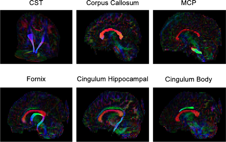
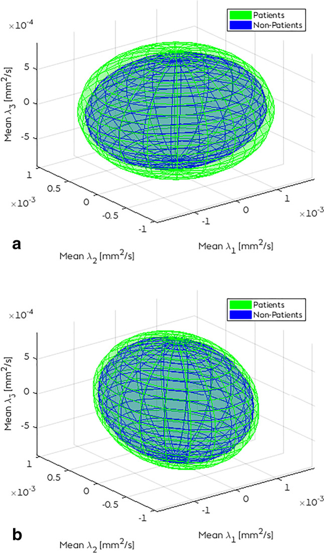
Methods: DTI datasets of 51 non-operated patients with syndromic craniosynostosis with a median [IQR] age of 0.40 [0.25] years were compared with 17 control subjects with a median of 1.20 [0.85] years. Major white matter tract pathways were reconstructed with ExploreDTI from MRI brain datasets acquired on a 1.5 T MRI system. Eigenvalues of these tract data were examined, with subsequent assessment of the affected tracts. Having syndromic craniosynostosis (versus control), gender, age, frontal occipital horn ratio (FOHR), and tract volume were treated as independent variables.
Results: ʎ2 and ʎ3 of the tracts genu of the corpus callosum and the hippocampal segment of the cingulum bundle show a ƞ2 > 0.14 in the comparison of patients vs controls, which indicates a large effect on radial diffusivity. Subsequent linear regressions on radial diffusivity of these tracts show that age and FOHR are significantly associated interacting factors on radial diffusivity (p < 0.025).
Conclusion: Syndromic craniosynostosis shows not to be a significant factor influencing the major white matter tracts. Enlargement of the ventricles show to be a significant factor on radial diffusivity in the tracts corpus callosum genu and the hippocampal segment of the cingulate bundle.
Clinical trial registration: MEC-2014-461.
Keywords: Craniosynostosis; DTI; Diffusion tensor imaging; Syndrome; Tractography.
© 2022. The Author(s).
Conflict of interest statement
All authors declare no conflict of interests.
Figures
References
-
- Britto JA, Evans RD, Hayward RD, et al (2001) From genotype to phenotype: the differential expression of FGF, FGFR, and TGFbeta genes characterizes human cranioskeletal development and reflects clinical presentation in FGFR syndromes. Plast Reconstr Surg 108:2026–2039; discussion 2040–2026 - PubMed
MeSH terms
Grants and funding
LinkOut - more resources
Full Text Sources
Research Materials
