Review
. 2023 Jan;475(1):37-48.
doi: 10.1007/s00424-022-02721-8. Epub 2022 Jun 28.

Could respiration-driven blood oxygen changes modulate neural activity?

Affiliations
Review

Could respiration-driven blood oxygen changes modulate neural activity?

Qingguang Zhang et al. Pflugers Arch. 2023 Jan.

Abstract

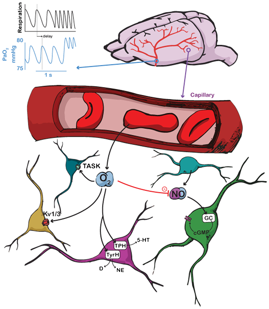
Oxygen is critical for neural metabolism, but under most physiological conditions, oxygen levels in the brain are far more than are required. Oxygen levels can be dynamically increased by increases in respiration rate that are tied to the arousal state of the brain and cognition, and not necessarily linked to exertion by the body. Why these changes in respiration occur when oxygen is already adequate has been a long-standing puzzle. In humans, performance on cognitive tasks can be affected by very high or very low oxygen levels, but whether the physiological changes in blood oxygenation produced by respiration have an appreciable effect is an open question. Oxygen has direct effects on potassium channels, increases the degradation rate of nitric oxide, and is rate limiting for the synthesis of some neuromodulators. We discuss whether oxygenation changes due to respiration contribute to neural dynamics associated with attention and arousal.

Keywords: Cognition; Neural excitability; Nitric oxide; Oxygen; Respiration.


Conflict of interest statement

Competing interests:

The authors have no competing interests to declare that are relevant to the content of this article.

Figures

Figure 1:
Figure 1:
Schematic showing different pathways by which oxygen can modulate neural excitability. Top, oxygen levels in major supply arteries oscillate on a breath-by-breath basis, as well as showing an overall increase with respiration rate. Scale is for expected values in a mouse. Respiration shows idealized measurement from a thermocouple, with upswings representing exhalation. Bottom left, oxygen modulates K+ channels and TASK activity in neurons. Bottom middle, oxygen modulates tryptophan hydroxylase (TPH) synthesis of serotonin (5-HT) and tyrosine hydroxylase (TyrH) synthesis of dopamine (D) and norepinephrine (NE). Coloration of neurons is aesthetic. Bottom right, oxygen decreases nitric oxide (NO) concentrations which modulates neural activity.
Figure 2
Figure 2
(a-b) Respiration drives changes in cerebral and blood oxygenation. (a) Measuring respiration using a thermocouple. Top, example data showing tissue oxygenation in the somatosensory cortex of an awake, headfixed mouse measured using an oxygen sensitive microelectrode (black trace) and respiratory rate (orange trace), during locomotion. Middle, signal from a thermocouple placed near the nostril of the mouse. The thermocouple voltage tracks inhalation and exhalation due to the higher temperature of exhaled air, which causes increase in the thermocouple signal. Bottom left, expanded thermocouple signal showing of the detection of the onset of inspiratory (magenta dot) and expiratory phase (blue dot). Bottom right, schematic showing respiration measurement using a thermocouple. (b) Example data showing the temporal relation between respiratory rate (black) and oxygen tension (PaO2, blue) in the center of one artery in somatosensory cortex of a mouse during periods of rest. The phase shift is caused by transit time from lungs to brain. (c) PaO2 fluctuates within the respiratory cycle. The PaO2 change in one artery of a headfixed, un-anesthetized mouse during the respiratory cycle at rest was measured using an intravascularly-injected phosphorescent oxygen dye using a two-photon microscope. This technique allows measurement of the concentration of oxygen in the blood plasma from a single location in the vasculature. PaO2 data (15 recordings with each of 50 seconds in duration) were aligned to the offset of inspiration. Each circle denotes averaged PaO2 over a short window (20 ms) and over the 15 recordings. The solid curve denotes filtering of data (first order binomial filter, 5 repetitions). Tmin denotes the time period (40 ms) PaO2 reaches minimum. Tmax denotes the time period (40 ms) PaO2 reaches maximum. (d) Example data showing the temporal relation between respiratory rate (black) and pupil diameter (blue, an indicator of noradrenergic activity) during periods of rest in an awake, headfixed mouse. (e) Cross-correlation between respiratory rate and pupil diameter during periods of rest. Gray shaded area indicates 95% confidence interval. (f-g) Nasal inhalation at visuospatial task onset is associated with improved performance in humans. (f) Mean event-related nasal respiratory signal used to trigger trial-onset time-locked to inhalation (orange) or exhalation (blue). Time 0 denotes task initiation. The grey rectangle along the x axis represents the stimulus (1,200 ms). Inset: a polar plot of the respiratory phase (in degrees) at trial onset is shown. The orange and blue bins are trials triggered by inhalation and exhalation, respectively (n = 28). (g) Scatter plot of performance in the EEG visuospatial task in inhalation and exhalation. Each point is a participant (n = 28). The diagonal line is the unit slope line (x = y). Thus, if points accumulate below the line, this means performance was better during inhalation. In the inlay, the mean group performance is shown. Error bars are SEM. a-e adapted from [125] , f-g adapted from [77].

Similar articles

Cited by

References

    1. Agvald P, Adding LC, Artlich A, Persson MG, Gustafsson LE (2002) Mechanisms of nitric oxide generation from nitroglycerin and endogenous sources during hypoxia in vivo. Br J Pharmacol 135:373–382. doi:10.1038/sj.bjp.0704489 - DOI - PMC - PubMed
    1. Amann M, Eldridge MW, Lovering AT, Stickland MK, Pegelow DF, Dempsey JA (2006) Arterial oxygenation influences central motor output and exercise performance via effects on peripheral locomotor muscle fatigue in humans. J Physiol 575:937–952. doi:10.1113/jphysiol.2006.113936 - DOI - PMC - PubMed
    1. Angelova PR, Kasymov V, Christie I, Sheikhbahaei S, Turovsky E, Marina N, Korsak a, Zwicker J, Teschemacher aG, Ackland GL, Funk GD, Kasparov S, Abramov aY, Gourine aV (2015) Functional Oxygen Sensitivity of Astrocytes. Journal of Neuroscience 35:10460–10473. doi:10.1523/JNEUROSCI.0045-15.2015 - DOI - PMC - PubMed
    1. Attwell D, Buchan AM, Charpak S, Lauritzen M, Macvicar BA, Newman EA (2010) Glial and neuronal control of brain blood flow. Nature 468:232–243 - PMC - PubMed
    1. Balestrino M, Somjen GG (1988) Concentration of carbon dioxide, interstitial pH and synaptic transmission in hippocampal formation of the rat. J Physiol 396:247–266. doi:10.1113/jphysiol.1988.sp016961 - DOI - PMC - PubMed

Publication types

LinkOut - more resources