

Giant cell tumor of soft tissue of the colon: a case report and review of the literature
- PMID: 35761215
- PMCID: PMC9235156
- DOI: 10.1186/s12876-022-02391-x
Giant cell tumor of soft tissue of the colon: a case report and review of the literature
Abstract
Background: A giant cell tumor (GCT) is a benign neoplasm characterized by mixture of mononuclear cells and multinucleated cells. A GCT of soft tissue (GCT-ST) is developed in various extraosseous sites, but GCT-ST of the gastrointestinal tract is very rare. GCT-ST usually has a benign course, but rare cases reported malignant potential of the tumor. Therefore, complete resection is required to prevent local recurrence or distant metastasis.
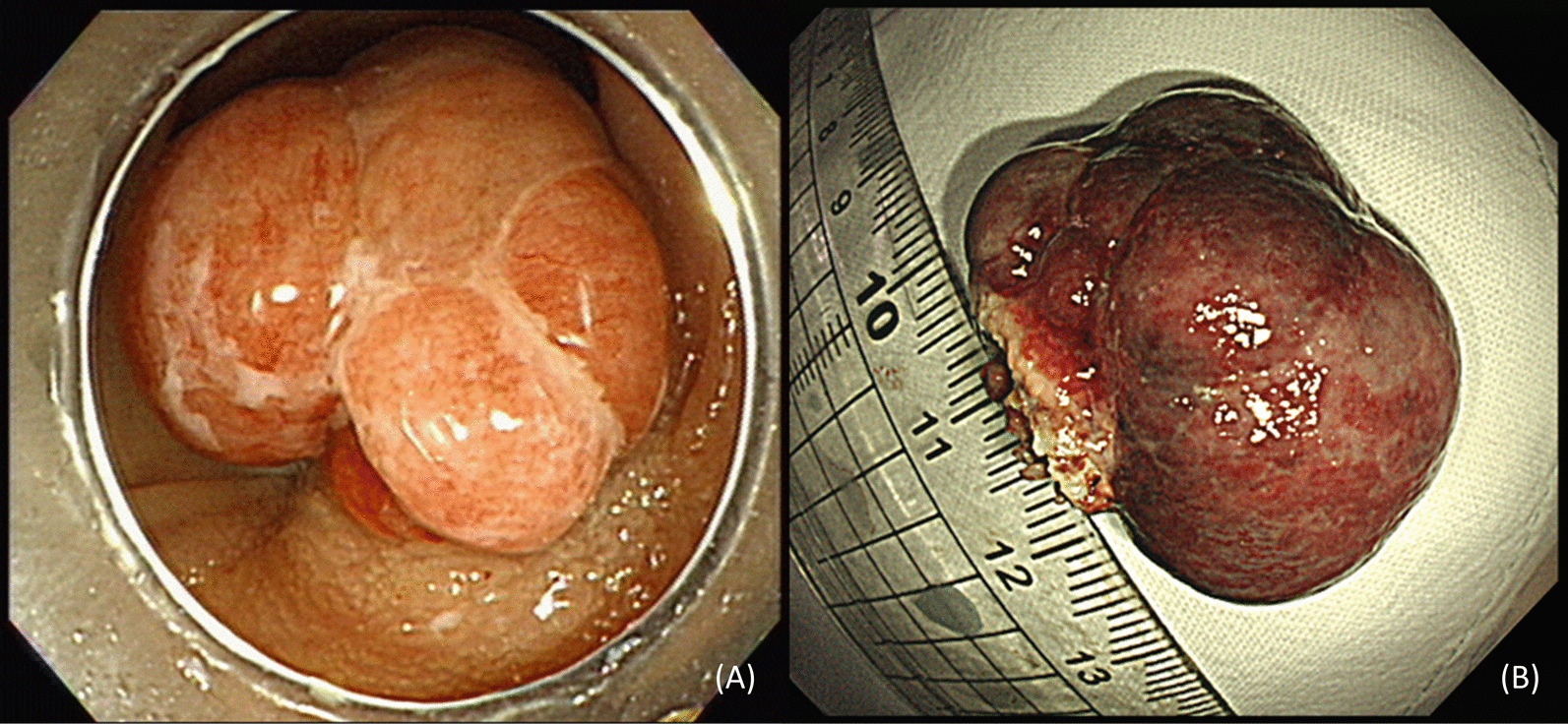
Case presentation: A 53-year-old woman was admitted for follow-up colonoscopy who underwent the colorectal endoscopic submucosal dissection (ESD) of a laterally spreading tumor at the hepatic flexure 6 months ago. A colonoscopy showed a polypoid mass about 3.5 × 2.5 cm at the previous ESD site. As endoscopic finding showed a smooth multi-nodular tumor without submucosal invasion, we performed endoscopic mucosal resection. Based on pathological and immunohistochemical findings, the lesion was diagnosed as a GCT-ST in the colon. Follow-up colonoscopy performed 6 months later revealed no evidence of recurrence.
Conclusion: This is the first report of a GCT-ST identified in the colon. Although GCT-ST generally has a benign clinical course, complete resection should be performed to prevent local recurrence and metastasis.
Keywords: Case report; Endoscopic mucosal resection; Giant cell tumors; Soft tissue neoplasm.
© 2022. The Author(s).
Conflict of interest statement
The authors declare that they have no competing interests.
Figures


Similar articles
-
Recurrent primary mediastinal giant cell tumor of soft tissue with radiological findings: a rare case report and literature review.World J Surg Oncol. 2017 Jul 26;15(1):137. doi: 10.1186/s12957-017-1205-5. World J Surg Oncol. 2017. PMID: 28747182 Free PMC article. Review.
-
Uterine giant cell tumor of soft tissue: A case report.Medicine (Baltimore). 2023 Oct 20;102(42):e35414. doi: 10.1097/MD.0000000000035414. Medicine (Baltimore). 2023. PMID: 37861517 Free PMC article.
-
Granular cell tumor of the cecum: Case report of mini invasive surgical resection and review of the literature.Int J Surg Case Rep. 2021 Oct;87:106397. doi: 10.1016/j.ijscr.2021.106397. Epub 2021 Sep 13. Int J Surg Case Rep. 2021. PMID: 34534816 Free PMC article.
-
Primary giant cell tumor of soft tissues: a study of 22 cases.Am J Surg Pathol. 2000 Feb;24(2):248-56. doi: 10.1097/00000478-200002000-00011. Am J Surg Pathol. 2000. PMID: 10680893
-
Giant Cell Tumor of Soft Tissue: A Rare Entity.Orthopedics. 2019 Jul 1;42(4):e364-e369. doi: 10.3928/01477447-20190624-04. Orthopedics. 2019. PMID: 31323108 Review.
Cited by
-
Unusual Multiple Recurrences of Soft Tissue Giant Cell Tumors in a Patient Above 60 Years.Cureus. 2024 Apr 28;16(4):e59195. doi: 10.7759/cureus.59195. eCollection 2024 Apr. Cureus. 2024. PMID: 38807802 Free PMC article.
References
-
- Icihikawa K, Tanino R. Soft tissue giant cell tumor of low malignant potential. Tokai J Exp Clin Med. 2004;29(3):91–95. - PubMed
Publication types
MeSH terms
LinkOut - more resources
Full Text Sources
Research Materials
Miscellaneous
