Review
. 2022 Jun 27;20(1):305.
doi: 10.1186/s12951-022-01510-w.

Pharmacokinetics of magnetic iron oxide nanoparticles for medical applications

Affiliations
Review

Pharmacokinetics of magnetic iron oxide nanoparticles for medical applications

Julia Nowak-Jary et al. J Nanobiotechnology. .

Abstract

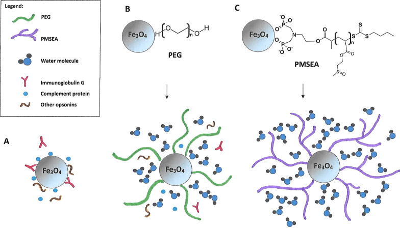
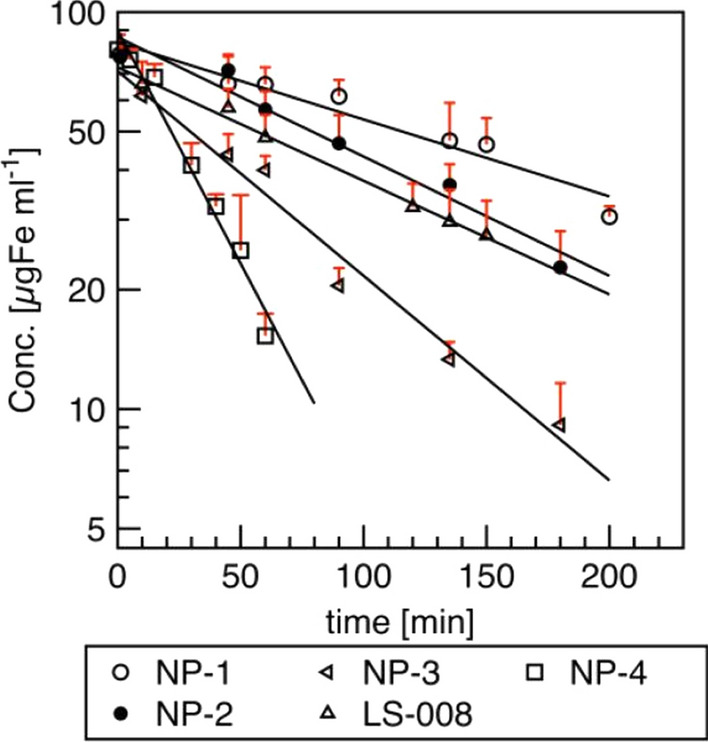
Magnetic iron oxide nanoparticles (MNPs) have been under intense investigation for at least the last five decades as they show enormous potential for many biomedical applications, such as biomolecule separation, MRI imaging and hyperthermia. Moreover, a large area of research on these nanostructures is concerned with their use as carriers of drugs, nucleic acids, peptides and other biologically active compounds, often leading to the development of targeted therapies. The uniqueness of MNPs is due to their nanometric size and unique magnetic properties. In addition, iron ions, which, along with oxygen, are a part of the MNPs, belong to the trace elements in the body. Therefore, after digesting MNPs in lysosomes, iron ions are incorporated into the natural circulation of this element in the body, which reduces the risk of excessive storage of nanoparticles. Still, one of the key issues for the therapeutic applications of magnetic nanoparticles is their pharmacokinetics which is reflected in the circulation time of MNPs in the bloodstream. These characteristics depend on many factors, such as the size and charge of MNPs, the nature of the polymers and any molecules attached to their surface, and other. Since the pharmacokinetics depends on the resultant of the physicochemical properties of nanoparticles, research should be carried out individually for all the nanostructures designed. Almost every year there are new reports on the results of studies on the pharmacokinetics of specific magnetic nanoparticles, thus it is very important to follow the achievements on this matter. This paper reviews the latest findings in this field. The mechanism of action of the mononuclear phagocytic system and the half-lives of a wide range of nanostructures are presented. Moreover, factors affecting clearance such as hydrodynamic and core size, core morphology and coatings molecules, surface charge and technical aspects have been described.

Keywords: Blood half-life; Endocytosis; Iron oxide magnetic nanoparticles; Pharmacokinetics.


Conflict of interest statement

The authors declared no potential conflicts of interest with respect to the research, authorship, and publication of this article.

The authors declare no potential conflicts of interest.

Figures

Fig. 1
Fig. 1
Magnetic nanoparticle internalization by opsonization and phagocytosis (A) and caveolin mediated endocytosis (CVME) (B)
Fig. 2
Fig. 2
Semiquantitative analysis of the protein corona composition of NP-DMSA and NP-PEG-(NH2)2(2000). Inmunoglobulins (A), Lipoproteins (B), Complement pathway (C), Transport (D), Acute phase (E), Coagulation (F). Republished from Ref. 31 under the terms of the Creative Commons Attribution Licence (CC BY) (http://creativecommons.org/licences/by/4.0/)
Fig. 3
Fig. 3
Delineation showing the size dependent physiological barriers against magnetic nanoparticles blood circulation. Even the smallest magnetic nanoparticles do not cross the blood-brain barrier and the blood vessel epithelium in the muscles, as long as the tissues are not cancerous (A). Nanoparticles with a diameter of about 5 nm or less are able to penetrate through small pores such as in the epithelium of the lungs and skin (B). Sinusoidal capillaries in the liver are fenestrated (100–180 nm) and lined with the Kupffer cells which quickly uptake large nanoparticles (> 100 nm) or agglomerates tagged with opsonins, whereas smaller nanoparticles (< 100 nm) are captured and hidden in the Disse space from where they can be collected by hepatocytes (C). Nanoparticles larger than about 200 nm get trapped in the marginal zones and the red pulp of the spleen, where they are absorbed by splenic macrophages (D). In the kidneys, nanoparticles with dH < 10–15 nm in diameter are filtered out, whereas nanoparticles with dH < 50–60 nm can penetrate through the pores in the intestines and glands (E)
Fig. 4
Fig. 4
The main non-spherical shapes of MNPs: nanorods (A), nanowires (B), nanotubes (C), nanodisks (D). SEM images A, B, D republished from Ref. 242, 243, 244, respectively, under the terms of the Creative Commons Attribution Licence (CC BY) (http://creativecommons.org/licences/by/4.0/); SEM image C republished from Ref. 245 with permission of Elsevier
Fig. 5
Fig. 5
The uptake of the MNPs by the macrophages is usually preceded by opsonization, which involves the attachment of specific proteins on the surface of the nanostructures (A). The „stealth” effect of the one of the most popular coating materials: PEG [poly(ethylene glycol) is explained by the high level of hydratation of the hydrophilic polyetherbackbone and its large conformational freedom, which causes the reduction of overall blood plasma protein adsorption and prevents MNPs agglomeration (B). Highly hydrophilic PMSEA [poly(2-(methylsulfinyl)ethyl acrylate] coating turned out to be even more resistant to protein binding as compared to PEG and thereby provides great low-fouling properties (C)
Fig. 6
Fig. 6
Clearance of MNPs samples from ex vivo Magnetic Particle Spectroscopy measurements; n = 3 per time point. Pharmacokinetic parameters were obtained after fitting data to a first-order elimination model. Republished from Ref. 79 under the terms of the Creative Commons Attribution Licence (CC BY) (http://creativecommons.org/licences/by/4.0/)
Fig. 7
Fig. 7
Interactions between positively (A–C) and negatively (D) charged magnetic nanoparticles (MNPs) and the plasma membrane. Electrostatic interactions with cationic MNPs and anionic syndecans and glypicans containing heparan sulfate (A). Nonspecific cationic MNPs interactions with anionic phospholipids (B). Transient pore formation by small cationic MNPs (≤ 20 nm) due to the strong attraction to the inner membrane layer in phosphatidylserine-rich regions (C). Local membrane gelation induced by anionic MNPs in phosphatidylcholine-rich membrane microdomains (D)
Fig. 8
Fig. 8
Time-dependent internalization profiles of CNP:Fe, P-CNP:Fe, and H-CNP:Fe internalized by J774A.1 macrophages (A). Comparison of intercellular Fe concentration of J774A.1 macrophages after 24 h incubation with different NPs (B). The CLSM images of J774A.1 macrophages after incubation for 24 h with different NPs (C). Republished from Ref. 209 with permission of Royal Society of Chemistry

Similar articles

Cited by

References

    1. Lu AH, Zhang XQ, Sun Q, Zhang Y, Song Q, Schüth F, et al. Precise synthesis of discrete and dispersible carbon-protected magnetic nanoparticles for efficient magnetic resonance imaging and photothermal therapy. Nano Res. 2016;9(5):1460–9. doi: 10.1007/s12274-016-1042-9. - DOI
    1. Zhou Q, Wei Y. For better or worse, iron overload by superparamagnetic iron oxide nanoparticles as a mri contrast agent for chronic liver diseases. Chem Res Toxicol. 2017;30(1):73–80. doi: 10.1021/acs.chemrestox.6b00298. - DOI - PubMed
    1. Yin PT, Shah BP, Lee KB. Combined Magnetic nanoparticle-based MicroRNA and hyperthermia therapy to enhance apoptosis in brain cancer cells. Small. 2014;10(20):4106–12. - PMC - PubMed
    1. Obaidat IM, Narayanaswamy V, Alaabed S, Sambasivam S, Muralee Gopi CVV. Principles of magnetic hyperthermia: a focus on using multifunctional hybrid magnetic nanoparticles. Magnetochemistry. 2019;5(4):67. doi: 10.3390/magnetochemistry5040067. - DOI
    1. Arruebo M, Fernández-Pacheco R, Ibarra MR, Santamaría J. Magnetic nanoparticles for drug delivery. Nano Today. 2007;2(3):22–32. doi: 10.1016/S1748-0132(07)70084-1. - DOI

LinkOut - more resources