

In vivo experimental validation of detection of gastric slow waves using a flexible multichannel electrogastrography sensor linear array
- PMID: 35761323
- PMCID: PMC9238032
- DOI: 10.1186/s12938-022-01010-w
In vivo experimental validation of detection of gastric slow waves using a flexible multichannel electrogastrography sensor linear array
Abstract
Background: Cutaneous electrogastrography (EGG) is a non-invasive technique that detects gastric bioelectrical slow waves, which in part govern the motility of the stomach. Changes in gastric slow waves have been associated with a number of functional gastric disorders, but to date accurate detection from the body-surface has been limited due to the low signal-to-noise ratio. The main aim of this study was to develop a flexible active-electrode EGG array.
Methods: Two Texas Instruments CMOS operational amplifiers: OPA2325 and TLC272BID, were benchtop tested and embedded in a flexible linear array of EGG electrodes, which contained four recording electrodes at 20-mm intervals. The cutaneous EGG arrays were validated in ten weaner pigs using simultaneous body-surface and serosal recordings, using the Cyton biosensing board and ActiveTwo acquisition systems. The serosal recordings were taken using a passive electrode array via surgical access to the stomach. Signals were filtered and compared in terms of frequency, amplitude, and phase-shift based on the classification of propagation direction from the serosal recordings.
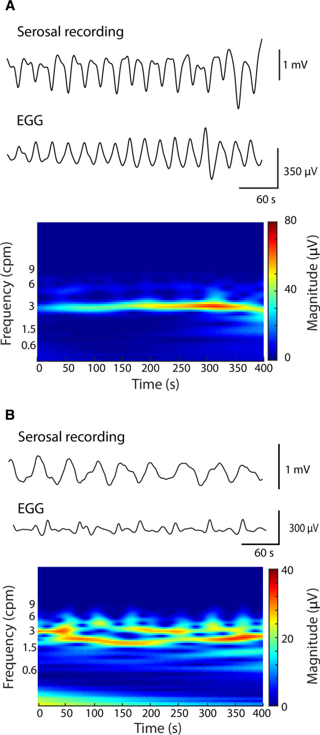
Results: The data were compared over 709 cycles of slow waves, with both active cutaneous EGG arrays demonstrating comparable performance. There was an agreement between frequencies of the cutaneous EGG and serosal recordings (3.01 ± 0.03 vs 3.03 ± 0.05 cycles per minute; p = 0.75). The cutaneous EGG also demonstrated a reduction in amplitude during abnormal propagation of gastric slow waves (310 ± 50 µV vs 277 ± 9 µV; p < 0.01), while no change in phase-shift was observed (1.28 ± 0.09 s vs 1.40 ± 0.10 s; p = 0.36).
Conclusion: A sparse linear cutaneous EGG array was capable of reliably detecting abnormalities of gastric slow waves. For more accurate characterization of gastric slow waves, a two-dimensional body-surface array will be required.
Keywords: EGG; Electrophysiology; Gastric slow waves; High-resolution mapping.
© 2022. The Author(s).
Conflict of interest statement
The authors declare that they have no competing interests.
Figures


Similar articles
-
Validation of noninvasive body-surface gastric mapping for detecting gastric slow-wave spatiotemporal features by simultaneous serosal mapping in porcine.Am J Physiol Gastrointest Liver Physiol. 2022 Oct 1;323(4):G295-G305. doi: 10.1152/ajpgi.00049.2022. Epub 2022 Aug 2. Am J Physiol Gastrointest Liver Physiol. 2022. PMID: 35916432
-
High-resolution electrical mapping of porcine gastric slow-wave propagation from the mucosal surface.Neurogastroenterol Motil. 2017 May;29(5):10.1111/nmo.13010. doi: 10.1111/nmo.13010. Epub 2016 Dec 29. Neurogastroenterol Motil. 2017. PMID: 28035728 Free PMC article.
-
What can be measured from surface electrogastrography. Computer simulations.Dig Dis Sci. 1997 Jul;42(7):1331-43. doi: 10.1023/a:1018869300296. Dig Dis Sci. 1997. PMID: 9246026
-
Electrogastrography: a document prepared by the gastric section of the American Motility Society Clinical GI Motility Testing Task Force.Neurogastroenterol Motil. 2003 Apr;15(2):89-102. doi: 10.1046/j.1365-2982.2003.00396.x. Neurogastroenterol Motil. 2003. PMID: 12680908 Review.
-
Current status of multichannel electrogastrography and examples of its use.J Smooth Muscle Res. 2013;49:78-88. doi: 10.1540/jsmr.49.78. J Smooth Muscle Res. 2013. PMID: 24662473 Free PMC article. Review.
Cited by
-
Electrogastrography measurement systems and analysis methods used in clinical practice and research: comprehensive review.Front Med (Lausanne). 2024 Jul 1;11:1369753. doi: 10.3389/fmed.2024.1369753. eCollection 2024. Front Med (Lausanne). 2024. PMID: 39011457 Free PMC article.
-
The effect of single and repeated doses of rivastigmine on gastric myoelectric activity in experimental pigs.PLoS One. 2023 Jun 1;18(6):e0286386. doi: 10.1371/journal.pone.0286386. eCollection 2023. PLoS One. 2023. PMID: 37262057 Free PMC article.
References
-
- Huizinga JD, Lammers WJEP. Gut peristalsis is governed by a multitude of cooperating mechanisms. Am J Physiol Gastrointest Liver Physiol. 2009;296(1):G1–8. - PubMed
-
- Schuffler MD. Chronic intestinal pseudo-obstruction syndromes. Med Clin North Am. 1981;65(6):1331–1358. - PubMed
-
- Camilleri M, Ford AC, Mawe GM, Dinning PG, Rao SS, Chey WD, et al. Chronic constipation. Nature Reviews Disease Primers: Nature Publishing Group; 2017. - PubMed
MeSH terms
LinkOut - more resources
Full Text Sources
