Review
. 2022 Jun 27;14(1):68.
doi: 10.1186/s13073-022-01075-1.

An introduction to spatial transcriptomics for biomedical research

Affiliations
Review

An introduction to spatial transcriptomics for biomedical research

Cameron G Williams et al. Genome Med. .

Abstract

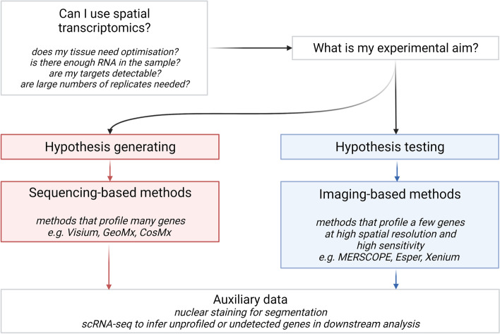
Single-cell transcriptomics (scRNA-seq) has become essential for biomedical research over the past decade, particularly in developmental biology, cancer, immunology, and neuroscience. Most commercially available scRNA-seq protocols require cells to be recovered intact and viable from tissue. This has precluded many cell types from study and largely destroys the spatial context that could otherwise inform analyses of cell identity and function. An increasing number of commercially available platforms now facilitate spatially resolved, high-dimensional assessment of gene transcription, known as 'spatial transcriptomics'. Here, we introduce different classes of method, which either record the locations of hybridized mRNA molecules in tissue, image the positions of cells themselves prior to assessment, or employ spatial arrays of mRNA probes of pre-determined location. We review sizes of tissue area that can be assessed, their spatial resolution, and the number and types of genes that can be profiled. We discuss if tissue preservation influences choice of platform, and provide guidance on whether specific platforms may be better suited to discovery screens or hypothesis testing. Finally, we introduce bioinformatic methods for analysing spatial transcriptomic data, including pre-processing, integration with existing scRNA-seq data, and inference of cell-cell interactions. Spatial -omics methods are already improving our understanding of human tissues in research, diagnostic, and therapeutic settings. To build upon these recent advancements, we provide entry-level guidance for those seeking to employ spatial transcriptomics in their own biomedical research.


Conflict of interest statement

CGW owns shares in 10X Genomics. The remaining authors declare that they have no competing interests.

Figures

Fig. 1
Fig. 1
Four different ways to record location and species of mRNA transcripts. Transcripts can be imaged directly in intact tissue by hybridization to fluorophore-labelled probes, or their locations can be recorded before they are extracted and undergo NGS. Transcript species can be imaged repeatedly with the same probes but different fluorophores to create a gene-specific barcode as in ISH. Short probes can also be imaged to read along an amplified transcript and determine its sequence as in ISS. Arrays of spatially barcoded probes can be used to label mRNAs with a sequence indicating location before undergoing NGS. Finally, cells or regions of interest can be directly microdissected and their locations recorded before their transcriptomes undergo NGS. Created with biorender.com
Fig. 2
Fig. 2
Design considerations for spatial transcriptomics experiments. Spatial transcriptomics technologies can satisfy a variety of experimental aims if the correct platform and design are chosen. Here, we have outlined a simple distinction between hypothesis testing—highly targeted experiments to examine regulation of defined genes and pathways—and hypothesis generation, which aims to reveal mechanisms without bias. Thus, we suggest hypothesis testing is best suited to efficient, targeted, and spatially-resolved ISH- and ISS-based methods. Conversely, hypothesis generation is best served by unbiased array- and microdissection-based methods that generate large volumes of data. However, researchers should note that there are a range of other considerations like the tissue type, quality of mRNA in the tissue, and amenability to generating a single-cell reference dataset that will also affect the choice of method or design
Fig. 3
Fig. 3
Typical structure of spatial transcriptomics analysis. Data are first preprocessed using technique-specific methods and algorithms. Normalization methods account for technical variation. Downstream analyses may be performed with a range of general-purpose transcriptomic analysis packages or with specialized methods for spatial transcriptomics. Created with biorender.com

References

    1. Tang F, et al. mRNA-Seq whole-transcriptome analysis of a single cell. Nat Methods. 2009;6(5):377–382. - PubMed
    1. Haque A, et al. A practical guide to single-cell RNA-sequencing for biomedical research and clinical applications. Genome Med. 2017;9(1):75. - PMC - PubMed
    1. Lacar B, et al. Nuclear RNA-seq of single neurons reveals molecular signatures of activation. Nat Commun. 2016;7:11022. - PMC - PubMed
    1. Svensson V, Vento-Tormo R, Teichmann SA. Exponential scaling of single-cell RNA-seq in the past decade. Nat Protoc. 2018;13(4):599–604. - PubMed
    1. Stubbington MJT, et al. T cell fate and clonality inference from single-cell transcriptomes. Nat Methods. 2016;13(4):329–332. - PMC - PubMed

Publication types

LinkOut - more resources