. 2022 Jun 27;15(1):23.
doi: 10.1186/s13072-022-00457-4.

Multiple epigenetic factors co-localize with HMGN proteins in A-compartment chromatin

Affiliations

Multiple epigenetic factors co-localize with HMGN proteins in A-compartment chromatin

Bing He et al. Epigenetics Chromatin. .

Abstract

Background: Nucleosomal binding proteins, HMGN, is a family of chromatin architectural proteins that are expressed in all vertebrate nuclei. Although previous studies have discovered that HMGN proteins have important roles in gene regulation and chromatin accessibility, whether and how HMGN proteins affect higher order chromatin status remains unknown.

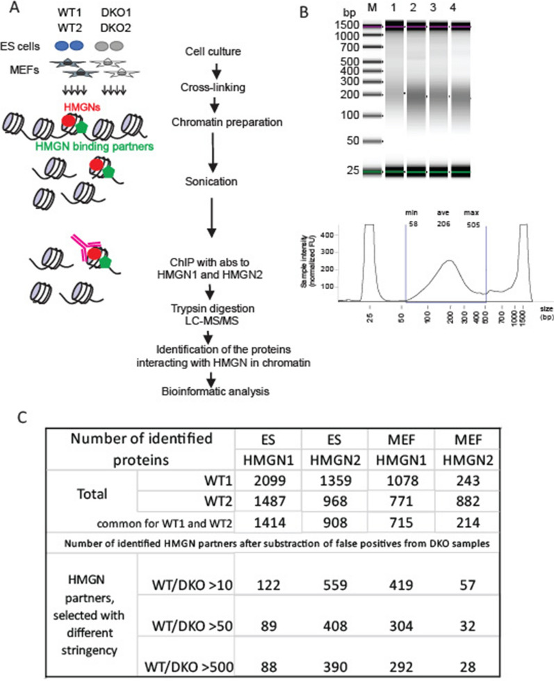
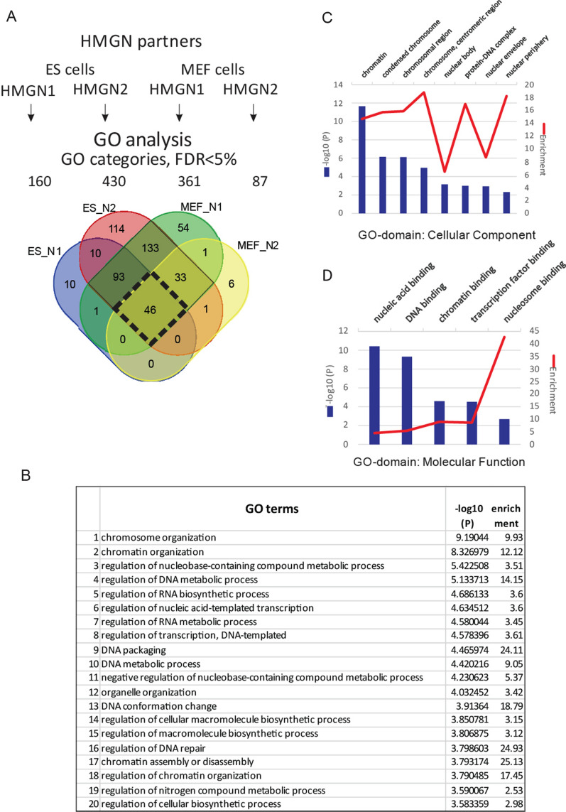
Results: We examined the roles that HMGN1 and HMGN2 proteins play in higher order chromatin structures in three different cell types. We interrogated data generated in situ, using several techniques, including Hi-C, Promoter Capture Hi-C, ChIP-seq, and ChIP-MS. Our results show that HMGN proteins occupy the A compartment in the 3D nucleus space. In particular, HMGN proteins occupy genomic regions involved in cell-type-specific long-range promoter-enhancer interactions. Interestingly, depletion of HMGN proteins in the three different cell types does not cause structural changes in higher order chromatin, i.e., in topologically associated domains (TADs) and in A/B compartment scores. Using ChIP-seq combined with mass spectrometry, we discovered protein partners that are directly associated with or neighbors of HMGNs on nucleosomes.

Conclusions: We determined how HMGN chromatin architectural proteins are positioned within a 3D nucleus space, including the identification of their binding partners in mononucleosomes. Our research indicates that HMGN proteins localize to active chromatin compartments but do not have major effects on 3D higher order chromatin structure and that their binding to chromatin is not dependent on specific protein partners.

Keywords: Chromatin structure; HMGN; Hi–C; Mass spectrometry.


Conflict of interest statement

The authors declare that they have no competing interests.

Figures

Fig. 1
Fig. 1
HMGN proteins (HMGN1 and HMGN2) are enriched at A compartments. A Percentage of HMGN1 and HMGN2 ChIP-seq peaks located in A compartment in MEFs, rBs, ESCs, and iPSCs. BE Left panels: average HMGN1 signals in 200 kb windows across boundaries from B compartment to A compartment in MEF, rB, ESC and iPSC cells. BE Right panels: individual examples of HMGN ChIP-seq signals enriched in A compartment and overlap with active enhancer maker H3K27ac. Snapshots are made from IGV genome browser
Fig. 2
Fig. 2
Unaltered 3D chromatin structure between WT and HMGN1/2 DKO cells. A Comparison of CScore of WT and DKO cells in MEF, rBs, and iPSCs. CScores are calculated at 25 kb resolution. B Examples of Juicebox illustration of similar Hi–C chromatin structure between WT and DKO at specific regions, at resolutions of 50 kb, 25 kb, and 10 kb in MEF, rB, and iPSCs. C Examples of similar TAD boundaries between WT and DKO in MEF, rB, and iPSCs. Hierarchical TADs are marked with lines of different colors
Fig. 3
Fig. 3
HMGN proteins bind to promoter interaction Regions (PIRs), which are highly enriched for cis-regulatory features involved in active transcription. A Chromatin features of promoter-interacting fragments detected with CHiCAGO. Yellow bars indicate overlaps of the genomic features with cis-interacting fragments within 1 Mb of promoter baits; blue bars indicate expected overlap values based on 100 random subsets of HindIII fragments. These subsets were selected to have a similar distribution of distances from gene promoters as the interacting fragments. Error bars represent 95% confidence intervals. Genomic features include ChIP-seq peaks of HMGN1/2, histone markers, CTCF, and p300. The difference between detected interactions and the expected value is significant for all genomic features with p value < 1e−30. The features involved in positive transcription regulation have a fold enrichment of ~ 2–3 while the repressive markers, H3K27me3 and H3K9me3, are either only slightly enriched or depleted. B Upper panel: snapshots of interactions with the Erc2 promoter identified with CHiCAGO in MEF cells. Lower panel: ChIP-seq signals of HMGN1, HMGN2 and H3K27ac in the same region. HMGN proteins specifically occupy at MEFs-specific site (Erc2) and its enhancers in MEF cells
Fig. 4
Fig. 4
Chromatin profiling by ChIP–MS identifies proteins that reside nucleosome-long proximity to HMGN-occupied regions of chromatin. A Schematic overview of ChIP–MS assay. Mouse embryonic stem cells and MEF cells were cross-linked, and the chromatin was isolated, sonicated, and immunoprecipitated. DKO cells served as a negative control. Overall, there were 16 samples: two immunoprecipitations (HMGN1 and HMGN2), two cell types (ES and MEF), two genotypes (WT and DKO), and two biological replicates. Proteins, co-purified with HMGN and identified by UHPLC/MS/MS, represent HMGN binding partners. B ChIP DNA visualized by the Agilent TapeStation System after the optimized sonication step. More than 70% of total DNA fragments ranged from shorter than 505 bp to longer than 58 bp. C The table with the total number of identified proteins in individual samples. The number of proteins selected as HMGN binding partners with various (WT/DKO) cutoff limits in various cells
Fig. 5
Fig. 5
Gene ontology analysis for the HMGN binding partners identified by chromatin proteomic profiling. A Venn diagram that shows the overlap between overrepresented GO categories (FDR > 5%) for HMGN1 and HMGN2 binding partners in both cell types. The black diamond shows the GO categories (46) in which proteins are overrepresented among both HMGNs and both cell types. B List of the top 20 out of 46 GO categories over-represented among both HMGNs and both cell types. C Cellular component GO-category domain analysis of HMGN1 and HMGN2 binding partners in both cell types. Top eight categories ranked by − log10 (P) value are depicted. Bar graphs − log 10 (P), P is FDR-corrected p value, blue and line graphs (enrichment score), red. D Molecular function GO category domain analysis of HMGN1 and HMGN2 binding partners in both cell types. Top five categories ranked by − log10 (P) value are shown

Similar articles

Cited by

References

    1. Woodcock CL, Ghosh RP. Chromatin higher-order structure and dynamics. Csh Perspect Biol. 2010;2(5):a000596. - PMC - PubMed
    1. Tremethick DJ. Higher-order structures of chromatin: the elusive 30 nm fiber. Cell. 2007;128(4):651–654. doi: 10.1016/j.cell.2007.02.008. - DOI - PubMed
    1. Ramani V, Shendure J, Duan Z. Understanding spatial genome organization: methods and insights. Genomics Proteomics Bioinformatics. 2016;14(1):7–20. doi: 10.1016/j.gpb.2016.01.002. - DOI - PMC - PubMed
    1. Szabo Q, Bantignies F, Cavalli G. Principles of genome folding into topologically associating domains. Sci Adv. 2019; 5(4). - PMC - PubMed
    1. Zheng H, Xie W. The role of 3D genome organization in development and cell differentiation. Nat Rev Mol Cell Bio. 2019;20(9):535–550. doi: 10.1038/s41580-019-0132-4. - DOI - PubMed

Publication types

LinkOut - more resources