. 2022 Jun 1;24(1):483.
doi: 10.3892/etm.2022.11410. eCollection 2022 Jul.

Fully endoscopic microvascular decompression for hemifacial spasm

Affiliations

Fully endoscopic microvascular decompression for hemifacial spasm

Hao Tian Jiang et al. Exp Ther Med. .

Abstract

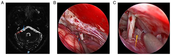
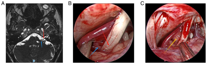
With the rapid development of endoscopic technology, fully endoscopic microvascular decompression (MVD) has been widely used in the treatment of hemifacial spasm (HFS), and has achieved good effect. The present study reviewed 5 cases of HFS treated by fully endoscopic MVD. After fully endoscopic MVD, the symptom of facial involuntary twitching was relieved in each of the 5 patients with an effective rate of 100%. Among the cases, 4 had no postoperative complications, such as cranial nerve dysfunction, and cerebellar or brainstem injury, while 1 patient had postoperative aseptic meningitis and recovered after follow-up treatment. In these 5 cases of MVD, endoscopy played an important role in identifying the offending blood vessels, which is of great significance to improve the surgical effect and safety. Furthermore, the postoperative effects showed that endoscopy has certain potential and advantages in MVD. Therefore, fully endoscopic MVD is also a feasible method for the treatment of HFS.

Keywords: endoscopy; hemifacial spasm; microvascular decompression.


Conflict of interest statement

The authors declare that they have no competing interests.

Figures

Figure 1
Figure 1
Patient 1. (A) Preoperative head magnetic resonance imaging of the patient. (B) The anterior inferior cerebellar artery and compressed cranial nerves were clearly visible under the endoscopic view. (C) The Teflon pad was placed between the root exit zone of cranial nerve 7 and the anterior inferior cerebellar artery under the endoscopic view. Red arrows indicate the compressed cranial nerve vessels, blue arrows indicate the anterior inferior cerebellar artery and the yellow arrow indicates the Teflon pad.
Figure 2
Figure 2
Patient 2. (A) Preoperative head magnetic resonance imaging of the patient. (B) The posterior inferior cerebellar artery and compressed cranial nerves were clearly visible under the endoscopic view. (C) The Teflon pad was placed between the root exit zone of cranial nerve 7 and the posterior inferior cerebellar artery under the endoscopic view. Red arrows indicate the compressed cranial nerve vessels, blue arrows indicate the posterior inferior cerebellar artery and the yellow arrow indicates the Teflon pad.
Figure 3
Figure 3
Patient 3. (A) Preoperative head magnetic resonance imaging of the patient. (B) In the first operation, the anterior inferior cerebellar artery and compressed cranial nerves were clearly visible under the endoscopic view. (C) The Teflon pad was placed between the root exit zone of cranial nerve 7 and the anterior inferior cerebellar artery under the endoscopic view in the first operation. (D and E) They are the images observed under the endoscope during the second operation, the offending blood vessel is the small artery. Red arrows indicate the compressed cranial nerve vessels, blue arrows indicate the offending blood vessels and yellow arrows indicate the Teflon pad.
Figure 4
Figure 4
Patient 4. (A) Preoperative head magnetic resonance imaging of the patient. (B) The the posterior inferior cerebellar artery and compressed cranial nerves were clearly visible under the endoscopic view. (C) The Teflon pad was placed between the root exit zone of cranial nerve 7 and the posterior inferior cerebellar artery under the endoscopic view. Red arrows indicate the compressed cranial nerve vessels, blue arrows indicate the posterior inferior cerebellar artery and the yellow arrow indicates the Teflon pad.
Figure 5
Figure 5
Patient 5. (A) Preoperative head magnetic resonance imaging of the patient. (B) The the anterior inferior cerebellar artery and compressed cranial nerves were clearly visible under the endoscopic view. (C) The Teflon pad was placed between the root exit zone of cranial nerve 7 and the anterior inferior cerebellar artery under the endoscopic view. Red arrows indicate the compressed cranial nerve vessels, blue arrows indicate the anterior inferior cerebellar artery and the yellow arrow indicates the Teflon pad.

References

    1. Blue R, Li C, Spadola M, Saylany A, McShane B, Lee JYK. Complication rates during endoscopic microvascular decompression surgery are low with or without petrosal vein sacrifice. World Neurosurg. 2020;138:e420–e425. doi: 10.1016/j.wneu.2020.02.142. - DOI - PubMed
    1. Jannetta PJ. Neurovascular compression in cranial nerve and systemic disease. Ann Surg. 1980;192:518–525. doi: 10.1097/00000658-198010000-00010. - DOI - PMC - PubMed
    1. Gardned WJ. Concerning the mechanism of trigeminal neuralgia and hemifacial spasm. J Neurosurg. 1962;19:947–958. doi: 10.3171/jns.1962.19.11.0947. - DOI - PubMed
    1. McLaughlin MR, Jannetta PJ, Clyde BL, Subach BR, Comey CH, Resnick DK. Microvascular decompression of cranial nerves: Lessons learned after 4400 operations. J Neurosurg. 1999;90:1–8. doi: 10.3171/jns.1999.90.1.0001. - DOI - PubMed
    1. Teton ZE, Blatt D, Holste K, Raslan AM, Burchiel KJ. Utilization of 3D imaging reconstructions and assessment of symptom-free survival after microvascular decompression of the facial nerve in hemifacial spasm. J Neurosurg: 1-8, 2019 (Epub ahead of print). - PubMed

LinkOut - more resources