Case Reports
. 2022 Jun 18:27:101625.
doi: 10.1016/j.ajoc.2022.101625. eCollection 2022 Sep.

Recurrent Vogt-Koyanagi-Harada disease presenting with diffuse orbital inflammation

Affiliations
Case Reports

Recurrent Vogt-Koyanagi-Harada disease presenting with diffuse orbital inflammation

Brandon Fram et al. Am J Ophthalmol Case Rep. .

Abstract

Purpose: To report diffuse orbital inflammation as a manifestation of recurrent inflammation in a patient with Vogt-Koyanagi-Harada (VKH) disease.

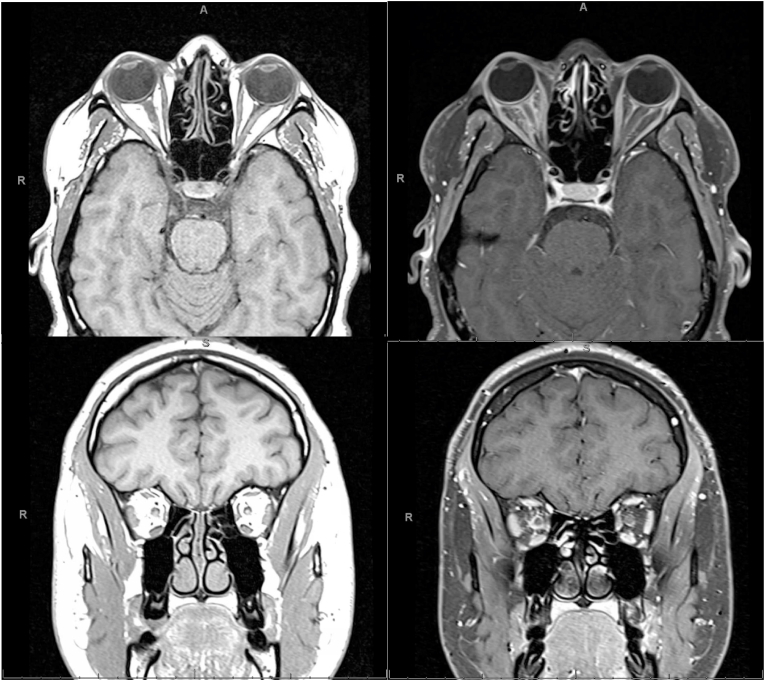
Observations: 20-year-old African American male, who was previously diagnosed with VKH, presented with right eye pain, swelling, and binocular double vision. He had run out of methotrexate while on steroid taper. Neuroimaging was consistent with diffuse orbital inflammation with myositis. He was started on intravenous steroids and then transitioned to oral steroids, with complete resolution of his symptoms.

Conclusions and importance: Central nervous system involvement as a manifestation of VKH has been previously reported, however, there have been no reports of orbital inflammatory syndrome resulting from VKH. Thus, in the appropriate clinical context, orbital signs may be recognized as features of recurrent VKH.

Keywords: Harada syndrome; Orbital inflammation; Uveomeningitis syndrome; VKH syndrome; Vogt-koyanagi-harada disease.


Conflict of interest statement

The following authors have no financial disclosures: BF, AF, VB, HZ.

Figures

Fig. 1
Fig. 1
Fundus photographs and optical coherence tomography, at the time of initial presentation and diagonis, demonstrating bilateral optic disc edema, choroidal folds, and sub-retinal fluid.
Fig. 2
Fig. 2
Color external photographs of patient in primary gaze, then performing ductions: up gaze, left gaze, down gaze, and right gaze. These photographs display the patient's right eye restrictions in extraocular movements during the first episode of orbital inflammation.
Fig. 3
Fig. 3
Axial and coronal T1-weighted fat saturated contrasted (left images) and post-contrast (right images) MRIs during patient's first episode of orbital inflammation. The post-contrast images display right-sided contrast enhancement and hyperintensity throughout the orbit, including intraconal fat. There is subtle right-sided enhancement of the extraocular muscles indicating myositis. Perineural enhancement is also seen in the post-contrast images. Additionally, there is mild right-sided proptosis. These findings of diffuse hyperintensity are indicative of an orbital inflammatory process.

References

    1. O'Keefe, Datoo Ghazala A., Rao Narsing A. Vogt-Koyanagi-Harada disease. Surv Ophthalmol. 2017;62:1–25. - PubMed
    1. Bonnet C., Daudin J.B., Monnet D., Brézin A. La maladie de Vogt-Koyanagi-Harada [Vogt-Koyanagi-Harada disease] J Fr Ophtalmol. 2017;40(6):512–519. - PubMed
    1. Lavezzo M.M., Sakata V.M., Morita C., et al. Vogt-Koyanagi-Harada disease: review of a rare autoimmune disease targeting antigens of melanocytes. Orphanet J Rare Dis. 2016;11:29. - PMC - PubMed
    1. Standardization of Uveitis Nomenclature (SUN) Working Group Classification criteria for vogt-koyanagi-harada disease. Am J Ophthalmol. 2021;228:205–211. - PMC - PubMed
    1. Baltmr Abeir, Lightman Sue, Tomkins-Netzer, Oren Vogt-Koyanagi-Harada syndrome - current perspectives. Clin Ophthalmol. 2016;10:2345–2361. - PMC - PubMed

Publication types

LinkOut - more resources